Leptothrix is a gram-negative anaerobic rod.
Refers to bacteria.
Has some affinity with mushrooms.
This is a representative of transit microflora.
Leptothrix often penetrates the genitourinary tract or oral cavity.
Can live there for a long time.
In this case, no pathological symptoms arise.
But symptoms are possible due to decreased immunity.
In this case, the disease is treated with antibacterial drugs.
Leptothrix in a smear - what is it?
Leptothrix can be detected in urogenital or oropharyngeal smears.
This bacterium can be seen under a microscope.
It consists of thin threads with segmented ends.
During microscopy, the laboratory technician notices long chains in the form of dots and dashes.
Leptothrix does not always cause inflammation.
But in some cases they are possible.
The bacterium exhibits pathogenicity most often in the presence of other microorganisms.
It intensifies inflammatory processes against the background of sexually transmitted infections.
It is also one of the causative agents of bacterial vaginosis.
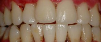
When it enters the oral cavity, leptothrix provokes inflammation of the gums, tonsils, and tongue.
May lead to darkening and destruction of tooth enamel.
What is Leptothrix
This is a micro-nanny that can exist and reproduce even without the presence of oxygen. Especially reproduces in conditions of high concentrations of carbon dioxide.
Leptotrix is a transitional form between bacteria and fungi. Viewed under a microscope, the appearance of fibers appears intertwined along the entire length of small dots.
Access to a woman's leptothrix body can pass through water from a natural tank or pool and is transmitted sexually.
Finding this infection in the back is not proof of a married spouse.
Causes of leptothrix
Leptothrix is very easy to become infected with.
The bacterium can spread from person to person through sexual contact.
But in men, leptothrix does not live in the genitourinary system.
Accordingly, partners of different sexes can become infected from each other only through oral sex.
However, after infection, the pathological process does not develop in most people.
Leptothrix exists peacefully on the mucous membrane of the genitourinary system.
It does not cause any problems for a long time.
Provoking factors are needed for bacteria to reach a large population and become pathogenic.
Such factors could be:
- decreased immunity;
- long-term uncontrolled antibiotic therapy (if those groups of drugs are used to which leptothrix is not sensitive);
- HIV;
- diabetes;
- bacterial vaginosis (leptothrix against the background of this disease is often found in association with other microorganisms);
- infection with sexually transmitted infections.
Leptothrix is not only transmitted from person to person.
It can also exist in the external environment for a long time.
Leptothrix can be contracted in a swimming pool or through poorly disinfected tap water.
How dangerous is leptothrix and where does it come from?
Long-term parasitism of the bacterium on the oral mucosa leads to infection of nearby tissues and the development of leptotrichosis sepsis. Often manifested in the form of oral stomatitis. Some experts consider leptotrichosis to be similar to HIV infection.
The disease was found in 5% of women undergoing gynecological tests - vaginal smears. Recent foreign studies have made it possible to determine the connection between leptotrichia and bacterial vaginosis (vaginal dysbiosis). The established connection suggests that leptothrix is one of the signs of asymptomatic vaginosis in women.
It is quite easy to catch this infectious pathogen. The microorganism is transmitted sexually and domestically: through the water of stagnant reservoirs, insufficiently clean tap water, dirty bed linen, towels, etc. That is, it is possible to infect a sexual partner with leptotrichosis during sex. It is advisable for both to undergo treatment.
There is an opinion that intrauterine devices are a common cause for the development of the disease.
Back to contents
Leptothrix in the mouth
Very often, leptothrix filaments are detected when taking a swab from the mouth.
This bacterium is normally present in the oral cavity during childhood.
It is detected in 40% of children under 1 year of age.
But adults should not have leptothrix in their mouths.
Because its presence provokes inflammatory processes and negatively affects the condition of the teeth.
Leptotrichosis usually develops against the background of immunodeficiency.
It is considered a disease close to opportunistic mycoses.
Often develops in HIV-infected people at the AIDS stage.
Also, leptothrix of the tongue can occur against the background of chronic diseases of the oral cavity and gastrointestinal tract.
In most cases, the patient himself does not feel anything when this disease occurs.
He has no complaints or complains of a slight burning sensation.
It is noted on the tongue and the inner surface of the cheeks.
Upon examination, the doctor finds areas of white or gray plaque.
They cover all structures of the oral cavity.
There are no obvious signs of inflammation.
Leptothrix clusters can be seen with the naked eye.
They look like small (pinhead-sized) white dots.
Plaque cannot be removed easily.
When scraped, the plaque is removed, but underneath it reveals inflamed, loose, sometimes bleeding mucosa.
Leptothrix: features of infection
If leptothrix is detected in a vaginal smear, then a woman may have a reasonable question.
What is it and where did this infection come from? In appearance, the pathogen has a shape resembling threads.
The infection can live not only in the vagina, but also in the mouth. Because it feels good in conditions of high CO2 concentration. In addition, pathogens are often inhabitants of natural bodies of water. If leptothrix is detected in vaginal smears, the doctor should conduct an additional examination. It is necessary to identify the possible presence of other dangerous infections.
If tests for concomitant infections show a negative result, then there is nothing dangerous. The infection may go away on its own even without treatment.
The disease is often found in pregnant women. As for men, they develop the disease very rarely; more often they are carriers of the infection. The characteristics of the pathogen are such that the bacterium can exist and is even capable of reproducing under anaerobic conditions (without the presence of oxygen). Microorganisms can be inhabitants of the vagina and oral cavity of healthy people.
When living conditions change, bacteria begin to multiply rapidly and become dangerous to humans. Treatment of the disease is carried out only if infected people make any complaints. Microorganisms exhibit maximum activity in the spring and autumn.
Clinical course of leptotrichosis
Oral leptotrichosis is difficult to treat.
The disease can last for months and years.
Especially in patients who have reduced immunity.
Sometimes after a course of antibacterial drugs, bacteria die.
All symptoms disappear.
But after some time, when taking the smear again, leptothrix was again detected.
This may be due to:
- re-infection;
- preservation of the source of infection in hard-to-reach areas of the oral cavity;
- no use of systemic therapy (only local treatment).
Leptothrix may spread to the pharynx.
In this case, the person complains of pain when swallowing.
Upon examination, the doctor may detect reddened mucous membranes.
Prevention
Simple rules allow you to prevent leptothrix:
- Comply with sanitary and hygienic standards and rules,
- Use filters that purify tap water
- Do not swallow water when swimming in open waters,
- Use a condom during sexual intercourse
- Before pregnancy, visit a gynecologist or antenatal clinic,
- Be examined by a female doctor at least once every six months.
Author: Tamm Irina Nikolaevna, MDDO, Tartu, Estonia © for izpp.ru
Leptothrix in gynecology
Leptothrix is often found in the urogenital system in women.
This bacterium lives in the vagina.
It may be part of an opportunistic flora.
But it is contained in small quantities.
In an ideal state of the vaginal biocenosis, the number of all anaerobic bacteria does not exceed 1% of their total number.
Among these anaerobes there is also a place for Leptothrix.
With such a small amount, these bacteria are not detected in a smear by microscopy and do not cause pathological processes.
Most often, leptotrichosis of the vagina is detected against the background of bacterial vaginosis.
The disease is manifested by discharge and the appearance of gray or white spots on the walls of the vagina.
The doctor discovers them during examination.
It is from these areas of the mucosa that smears are taken, in which leptothrix is detected.
An increase in the number of anaerobes can be caused by:
- taking antibiotics
- decreased immunity
- suffered serious illnesses
- lack of sufficient vaginal hygiene, or, on the contrary, excessive hygiene with frequent use of detergents containing antibacterial components
When lactobacilli die, anaerobic microorganisms take their place.
The vaginal pH shifts.
Lactic acid and hydrogen peroxide are not produced.
Therefore, the female genitourinary system becomes vulnerable to infections.
A woman easily becomes infected with sexually transmitted diseases through sexual intercourse.
Nonspecific inflammation is often associated.
Bacterial vaginosis associated with Leptothrix occurs without treatment for years.
Self-healing is unlikely.
Anaerobes occupy most of the biocenosis and do not allow lactobacilli to recover.
Therefore, only antibacterial treatment helps to normalize the composition of the vaginal microflora.
After it is carried out, probiotics are prescribed to populate the vaginal environment with “useful” microbes.
Consequences and possible complications of leptotrichosis
It is important to understand that the presence of leptothrix in tests may indicate the possibility of developing an inflammatory process provoked by an STI. A relationship has also been established between opportunistic flora and bacterial vaginosis.
It is necessary to undergo a timely examination and the necessary course of treatment. Otherwise, the inflammatory process can provoke various negative consequences. As a result, the following complications may develop:
- Infection of the fetus and membranes, if the disease is detected during pregnancy and the pathology has not been treated.
- Congenital developmental anomalies in a newborn.
- An inflammatory process of the genitals, eyes, and oral cavity in a newborn, caused by infection during the passage of the baby through the mother’s birth canal.
- Tooth decay caused by pathological changes in the enamel due to the localization of the inflammatory process in the mouth.
Tests for leptothrix
Leptothrix is not at all difficult to detect.
This bacterium is visible under microscopy.
In most cases, targeted testing for leptotrichosis is not carried out.
The pathogen is identified during a bacterioscopic examination.
A swab is taken from the mouth or vagina of women.
Gram-negative rod-shaped bacteria are found in the clinical material.
They form chains in the form of alternating dots and dashes.
In turn, the detection of leptothrix is the reason for a more in-depth examination of the patient.
Because the presence of this microorganism is an indicator of other diseases.
Against the background of leptotrichosis, the following are very often detected:
1. Sexual infections.
In approximately 60% of cases, along with leptothrix, a thorough examination of the patient reveals candida, ureaplasma, trichomonas, chlamydia or other pathogens.
Therefore, all women diagnosed with leptotrichosis are prescribed a smear from the urethra, vagina and cervix for sexually transmitted diseases.
PCR is mainly used for diagnosis.
2. Bacterial vaginosis.
It can be diagnosed by microscopy alone, which is supplemented by an amino test.
Bacterial vaginosis is said to occur when leptothrix and other anaerobic flora significantly outnumber lactobacilli.
Vaginal dysbiosis is considered a condition when the number of lactobacilli is less than 90%.
To clarify the state of the biocenosis, seeding or PCR may be prescribed.
3. Immunodeficiency states.
Leptothrix is considered an indicator of reduced immunity.
This is because in most patients, Leptothrix does not cause symptoms and does not multiply to the point of being detected by microscopy.
If this happens, it is likely that the immune system has failed.
It cannot inhibit the proliferation of the microorganism.
Therefore, patients diagnosed with leptothrix are often tested for HIV.
All patients are recommended to consult an immunologist.
The doctor may order an immunogram.
Based on this, the presence or absence of immunodeficiency is determined.
If necessary, immunocorrection is carried out in the future.
Occasionally, PCR or tank culture is used to detect leptothrix.
Pharyngomycosis. diagnosis, prevention and treatment
N.L. KUNELSKAYA
, Doctor of Medical Sciences, Professor,
G.N.
IZOTOVA , Ph.D.,
V.Ya.
KUNELSKAYA , Doctor of Medical Sciences, Professor,
G.B.
SHADRIN , Ph.D.,
D.I.
KRASNIKOVA ,
O.A.
ANDREENKOVA ,
State Budgetary Healthcare Institution “MNPC of Otorhinolaryngology named after. L.I. Sverzhevsky" of the Moscow Department of Health. An analysis of the prevalence of pharyngomycosis was carried out based on scientific literature and the results of our own observations of 2,987 patients who sought help at the advisory department of the Center in the period 2008–2011.
The proportion of fungal infection of the ENT organs in the structure of chronic inflammatory pathology is shown: the proportion of fungal infection of the pharynx with pharyngitis increased to 29%, with chronic tonsillitis – to 17.2%. The spectrum of pathogens causing fungal infections of the pharynx was determined and analyzed. An increase in the proportion of Candida non albicans strains in the development of chronic inflammation was revealed. A proportion of aspergillus lesions of the mucous membranes of the pharynx and larynx, and palatine tonsils was revealed. A modern diagnostic and treatment scheme is proposed. In medicine, the problem of mycotic diseases is currently acquiring important social significance due to a significant increase in their frequency. Increased incidence of mycoses in the middle of the 20th century. is associated with many factors, primarily with the introduction of antibacterial drugs into medical practice, which undoubtedly increased the number of not only candidiasis, but also mycoses caused by filamentous fungi. The increase in the number of mycosis is facilitated by cytostatics and other drugs with immunosuppressive properties, as well as a combination of other reasons associated with the urbanization of modern life of the population [4, 5, 10, 14].
Currently, more than 400 species of fungi are potential causative agents of mycoses. The incidence of deep mycoses has also increased significantly, which includes some mycotic lesions of the ENT organs [2, 3, 10, 14].
The objective of this research was to identify the prevalence of fungal diseases of the pharynx and larynx, to determine the epidemiological characteristics of the causative agents of mycoses depending on the location of the inflammatory process, and to develop a treatment regimen for pharyngomycosis.
The presented results are an analysis of epidemiological clinical and laboratory studies conducted at the Moscow Scientific and Practical Center of Otorhinolaryngology in the period from 2008 to 2011 inclusive. Over these years, 2,987 patients of both sexes with chronic pharyngitis aged from 16 to 86 years were examined.
In addition to the mandatory clinical examination of patients with this pathology, mycological studies were also carried out. Mycological diagnostics were carried out as follows: all patients had their ENT organs examined using magnifying optical technology, endomicroscopy (pharyngomicroscopy, etc.), which made it possible to take samples of pathological material directly from the site of inflammation (Fig. 1) [2, 7, 11].
The first stage of research included mandatory microscopy of the obtained material in the form of preparations stained using the Gram method and calcofluor white. This method allows not only to identify fungi, but also to determine the presence of their active forms, for example, the formation of pseudomycelium. The calcofluor white staining method is based on the ability of this dye to form a stable bond with the polysaccharides of the cell wall of fungi and fluoresce under fluorescent lighting with a wavelength of 340 nm.
The second stage of research involved the mandatory inoculation of pathological discharge on elective media, which made it possible to carry out species characteristics of fungi. We used liquid Sabouraud medium to transport the collected samples. For inoculations, Sabouraud agar with chloramphenicol, Czapek–Dox agar, and chromogenic HiCrome Candida Differential Agar were used. If necessary, histological and immunological studies were performed. For species identification of isolated yeast-like fungi of the genus Candida, ID 32C test strips from BioMeriex were used. To record the results, a miniAPI device from BioMeriex was used. In some cases, they were limited to the use of chromogenic agar, which makes it possible to determine the types of fungi of the genus Candida: C. albicans, C. tropicalis, C. krusei, C. glabrata and Candidaspp.
As a result of such an examination of 2,987 (100%) patients, the mycotic nature of the disease was identified in 750 patients, i.e. 25.1%. These figures indicate the great importance of fungi as an infectious etiological factor in chronic inflammatory diseases of the pharynx and larynx, i.e., almost every fourth patient with this pathology of the ENT organs is diagnosed with mycosis. Among 2,987 patients with chronic pharyngitis and chronic inflammation of the tonsils, two groups were separately identified: Group 1 – 1,989 patients with various forms of chronic pharyngitis, of which mycosis was detected in 578 (29%): Group 2 – 998 patients with chronic inflammation palatine and/or pharyngeal tonsils, of which mycosis was detected in 172 cases (17.2%). The distribution of patients is presented in Table 1.
Thus, analysis of the results of our clinical and laboratory studies showed a large role of fungal pathology among patients suffering from chronic pharyngitis and tonsillitis.
Information about the infectious pathogens themselves of fungal infections of the ENT organs is of both scientific and practical interest. Knowledge of the epidemiology of fungal infection in diseases of the ENT organs is important, because it forces us to improve methods for their diagnosis and specific antifungal therapy.
Our laboratory mycological studies made it possible to identify the main causative agents of mycotic diseases of the pharynx and larynx. During cultural diagnostics of isolated fungi from the pathological focus, their generic and species affiliation was determined. As a result, it was established that the causative agents of pharyngomycosis in 750 patients in the vast majority of cases (97%) were yeast-like fungi of the genus Candida. Molds of the genus Aspergillus were isolated in 3% of patients (21 observations). This must be taken into account when carrying out treatment. The causative agents of mycosis of the pharynx and larynx are presented in Table 2.
Pharyngomycosis is not a highly contagious disease. Most often, inflammation of the mucous membranes caused by fungi of the genus Candida is secondary. Against the background of predisposing factors, fungi that saprophyte in the body may appear. The exception is molds, whose spores are widespread in the environment.
Depending on the degree of damage, pseudomembranous, hyperplastic, granulomatous and erosive-ulcerative clinical forms of pharyngomycosis are distinguished.
When studying epidemiological and pathogenetic aspects, a direct connection was established between taking modern antibacterial agents in doses exceeding therapeutic ones and the development of pharyngomycosis. It is also necessary to take into account self-treatment of pharyngeal diseases and improper care of removable dentures carried out by patients. Another predisposing factor is trauma to the mucous membrane, including instrumental, “high reflux” in gastoesophageal reflux disease, smoking [1, 6, 8].
The development of mycosis is also provoked by severe somatic diseases, for example, diabetes mellitus, blood diseases, malignant neoplasms, bronchial asthma.
Secondary immunodeficiency, which develops not only against the background of HIV infection, but also with long-term use of corticosteroids at a dose exceeding 0.5 mg/kg/day for more than 28 days, or other long-term immunosuppressive therapy significantly increases the risk of developing superficial mycosis. Staying in intensive care units for more than 10 days, prolonged tracheal intubation, and rare changes of the tracheostomy tube significantly contribute to the development of fungal infections of the pharynx and larynx.
Of scientific and practical interest are 21 observations of pharyngomycosis caused by fungi of the genus Aspergillus. This is due to the fact that most mycologists believed that fungal infections of the pharynx are caused only by fungi of the genus Candida albicans [1, 2, 6, 10, 12], as a result of which we carried out mycological studies in this group of patients especially carefully. These results were obtained with repeated repeated cultures of discharge from inflammation. All patients underwent a comprehensive examination, which included X-ray examinations of the lungs and paranasal sinuses, and no foci were identified in the lungs and paranasal sinuses from which the fungal infection could spread. In fungal aspergillus pharyngitis, the focus was located in the tissue of the palatine tonsils. In all patients, the clinical picture, confirmed by mycological studies, normalized after complex combined treatment, which included local and systemic effects on the focus of the fungal infection.
Among yeast-like fungi (n = 729) (Table 2), the most pathogenic fungus remains Candida albicans, which we identified in 223 patients (30.5%), in second place is Candida tropicalis - 52 observations (7.1%), in third place Candida krusei – 36 observations (5%), C.pseudotropicalis – 25 observations (3.5%). Mycosis of the ENT organs was also caused by other species of Candida: C.sake, C.parapsilosis, C.dubliniensis, C.ciferrii, C.hellermanii, C.holmii, C.glabrata, C.globosa, Candida spp.
The main criterion for diagnosing a fungal infection, in addition to complaints and clinical signs, is laboratory confirmation, and the titer of the isolated fungi must be at least 1.0 x 10⁴ CFU, or actively vegetating fungi must be isolated in smears.
In addition to cultural diagnostics, it is necessary to carry out a differential diagnosis of fungal infection of the pharynx and larynx with diseases such as leptotrichosis, lichen planus, true leukoplakia, soft leukoplakia, geographic tongue, tuberculosis, syphilis, malignant neoplasms, granulomatosis, pemphigus, rhomboid glossitis, Fordyce glands and etc.
Our epidemiological clinical and laboratory studies generally showed the widespread prevalence of fungal infections of the pharynx.
The next task was to develop a treatment regimen for fungal infection of the pharynx. We began treatment with elimination of the pathogen. Correction of general and local predisposing factors was carried out in parallel or in the second stage.
Therapy can be local and/or systemic. When carrying out treatment, it is necessary to take into account previous episodes of fungal infection and treatment with antifungals.
When carrying out treatment, we always started with topical medications. These include aqueous suspensions of polyene antimycotics and imidazole derivatives, prescribed in the form of aerosols, solutions, drops, and chewable tablets.
Local polyene antimycotics (nystatin, levorin, natamycin, amphotericin) in our country, unfortunately, are presented in forms that are not suitable for application to the mucous membrane of the oral cavity and pharynx, so the doctor and patient need to adapt them independently.
It is necessary to explain to the patient that any drug for local treatment should remain in the oral cavity for as long as possible. A nystatin tablet in the form of a paste, chewed after a meal, should remain in the mouth for several minutes. You can prepare a suspension of nystatin, for which the tablet is kneaded and mixed with a small volume of water.
An effective and convenient alternative is an imidazole derivative, clotrimazole, in the form of a glycerin-based solution for lubricating the mouth and pharynx. On our market it is represented by the drug Candide, which is a 1% solution of clotrimazole. The antimycotic effect of Candida is associated with a disruption in the synthesis of ergosterol, which is part of the cell membrane of fungi, which causes a change in its structure and properties and leads to cell lysis. Dermatophytes and yeast-like fungi of the genus Candida, Torulopsis glabrata, Rhodotorula are sensitive to clotrimazole. The drug also has an antimicrobial effect against gram-positive (staphylococci and streptococci) and gram-negative bacteria.
We recommend applying the solution to the affected areas of the oropharynx five times a day. In this case, you should make sure that all affected areas are treated. Improvement usually occurs on the 3rd–5th day of therapy, but use of the drug must be continued until the clinical manifestations of the disease are completely eliminated. Candide is usually well tolerated.
It is also possible to use solutions of antiseptics and dyes, however, these drugs are inferior in effectiveness to antimycotics, their continuous use causes irritation of the mucous membrane, and the pathogens of pharyngomycosis quickly develop resistance to them. Therefore, it is recommended to alternate local antiseptics every week. Antiseptic solutions should not be swallowed, unlike antimycotic solutions [10, 13].
Systemic drugs are not recommended as first choice and are prescribed only for specific indications. These are: ineffectiveness of local therapy, frequent relapses after local therapy, chronic perennial recurrent forms of fungal infection of the pharynx and larynx, immunodeficiency due to HIV infection, cancer, severe forms of diabetes, combination with candidiasis of smooth skin and its appendages. The drug of choice when prescribing systemic therapy to immunocompetent patients is fluconazole at a dose of 50–100 mg/day for 14 days. For the treatment of exacerbations of chronic pharyngomycosis in patients with immunodeficiency, the dose of fluconazole is increased to 200 mg/day, and the duration of therapy is up to 28 days, while it is mandatory to take an “anti-relapse dose” - 100 mg of fluconazole three times a week for life. Reserve drugs for systemic therapy of pharyngeal mycosis are itraconazole solution, used at a dose of 200 mg/day; posaconazole suspension 400 mg twice daily for the first 3 days, then 400 mg/day for 28 days. Alternative drugs are: amphotericin B intravenously at a dose of 0.3 mg/kg/day; voriconazole 6 mg/kg/day in two doses, then 4 mg/kg in two doses; caspofungin intravenously 70 mg on the first day, then 50 mg/day in one injection.
In general, treatment of pharyngeal mycosis in patients without immunodeficiency averages 2–3 weeks and should continue for 1 week after complaints and clinical signs of the disease have subsided.
There are also natural protection factors against fungi. These are intact, normally functioning border tissues - the mucous membrane; factors of specific and nonspecific immunity (lysozyme, secretory immunoglobulins - sIgA); the presence of representatives of normal microflora - the main antagonists of fungi (colonization immunity). The latter include probiotic bacterial complexes, for example Lorodent, which includes the Streptococcus salivarius K12 strain and 5 lactobacilli strains. Streptococcus salivarius K12 is a representative of the normal oral flora and forms colonization immunity in healthy people through the production of antibiotic substances. Our experience with the use of this probiotic complex has shown that the treatment period can be shortened, and the proportion of disease relapse has almost halved [9].
In general, the results of our research determine the scientific and practical importance of comprehensive coverage of this problem among otorhinolaryngologists, mycologists, laboratory doctors and doctors of other specialties.
Literature
1. Akulich N.I., Lopatin A.S. Fungal disease of the pharynx. Attending doctor. – 2003. No. 8. – pp. 34–37. 2. Arabian R.A., Klimko N.N., Vasilyeva N.V. Diagnosis of mycoses. A manual for doctors. St. Petersburg, 2004. – 185 p. 3. Klimko N.N. Mycoses: diagnosis and treatment. Guide for doctors. 2nd ed., revised. and additional – M.: VG Group, 2008. – 336 p. 4. Kryukov A.I., Kunelskaya V.Ya., Shadrin G.B. Epidemiology of fungal diseases of the upper respiratory tract and ear. Problems of medical mycology. St. Petersburg, 2011. T. 13, No. 1. – pp. 28–31. 5. Kryukov A.I., Turovsky A.B., Shadrin G.B. Mycoses in otorhinolaryngology. Journal of evidence-based medicine for practitioners “Consiliummedicum”. 2004. T. 6, No. 4. – P. 56. 6. Kubanova A.A., Potekaev N.S., Potekaev N.N. Guide to practical mycology - M., 2001. 144s. 7. Kunelskaya V.Ya. Mycoses in otorhinolaryngology. – M.: Medicine, 1989. – 320 p. 8. Kunelskaya V.Ya. Kasymov K. et al. Mycoses of the oropharynx. Guidelines. M., 1989. – 15 p. 9. Kryukov A.I., Kunelskaya N.L., Kunelskaya V.Ya., Gurov A.V. and others. New opportunities in the treatment of pharyngitis. Method. rec. M., 2013. – 12 p. 10. Sergeev A.Yu., Sergeev Yu.V. Fungal infections. Guide for doctors. 2nd ed. – M.: BINOM Publishing House, 2008. – 408 p. 11. Starosvetsky B.M. Chronic pharyngitis of fungal etiology. Vestn. otorhinol. – 1988. No. 5. – 27–31. 12. Blyth, CC Antifungal therapy in children with invasive fungal infections: a systematic review. / CC Blyth, P. Palasanthiran, TA O'Brien // Pediatrics 119 (2007) 772–784. 13. Gonzalez GM, Fothergill AW, Sutton DA, et al. In vitro activities of new and established triazoles against opportunistic filamentous and dimorphic fungi. Med Mycol 2005; 43:281–84. 14. Kaieda, S. Fungal infection in the otorhinolaryngologic area. / S. Kaieda // Nippon Rinsho. 2008 Dec; 66(12): 2290 - 3.
Leptothrix during pregnancy
Leptothrix rarely causes complications during pregnancy.
Nevertheless, such cases do occur.
There is now evidence that leptotrichosis increases the risk of:
- chorioamnionitis
- spontaneous abortion
- birth of underweight children
- bacteremia of newborns
The likelihood of postpartum endometritis increases.
Immunosuppressed women may develop sepsis after childbirth.
Some of these complications develop as a result of the direct pathogenic effect of Leptothrix.
Some are associated with decreased immunity.
In this case, leptothrix is not so much a cause as a marker.
After all, the presence of this bacterium indicates that not everything is in order with the woman’s immune system.
Leptothrix: preventive measures
Infection of women with Leptothrix, unlike men, occurs much more easily. And the process of getting rid of the infection is quite complex and lengthy. Therefore, to avoid infection with a microorganism, it is enough to adhere to certain rules. Strict adherence to safety measures will avoid not only infection, but also possible subsequent problems. Rules to help prevent Leptothrix infection:
- Careful adherence to personal hygiene requirements.
- For drinking, use filtered tap or bottled water.
- After swimming in ponds, visiting pools, saunas, hygiene procedures are carried out.
- Carrying out hygienic procedures at least twice a day with the mandatory use of special care products for intimate areas.
- When swimming, avoid getting water into your mouth.
- Regular visits to the gynecologist to select contraceptives. If an IUD is used, it is necessary not only to remove the device in a timely manner, but also to regularly take smears.
- Get tested for STDs at least once a year.
Visit a gynecologist every six months for a routine examination and pap smears.- Be sure to visit a woman's doctor at the stage of pregnancy planning in order to be examined for the presence of dangerous infections.
- In the absence of a regular sexual partner, the use of a condom is mandatory.
- Washing your hands after every sexual intercourse.
- If the test results show the presence of infection, then you must strictly follow the doctor’s instructions and take treatment seriously.
If you follow all these rules, you can avoid infection with Leptothrix.
Also prevent re-infections.
Leptothrix in men
A woman cannot infect a man with leptotrichosis through vaginal intercourse.
This bacterium is not adapted to live in the male urethra.
Therefore, men can have sex without fear with a woman who has vaginal leptotrichosis.
However, you should be aware of the possibility of infection from other bacteria.
After all, leptothrix often talks about bacterial vaginosis.
And with this disease, many other anaerobes are present in the vagina.
Some of them can cause anaerobic balanoposthitis in a man.
It occurs mainly in people with immunodeficiency.
The most common pathogen is gardnerella.
In addition, leptothrix is often detected in those who suffer from sexually transmitted diseases that occur in a latent form.
Therefore, a man can become infected with trichomoniasis, ureaplasmosis, chlamydia or other pathology.
For these reasons, it is better to avoid unprotected sexual contact with a woman until she is fully examined and treated.
In addition, a man's leptothrix can live in his mouth.
Therefore, it can become infected through oral sex.
At the same time, he himself also becomes a source of infection for his partners.
Symptoms and stages of liver cirrhosis
As liver cirrhosis progresses, it goes through three stages:
- Initial. Moderate fibrosis is practically asymptomatic. The disease becomes an accidental discovery during periodic medical examinations, medical examinations, and when examining a patient with related pathology.
- Clinical. Severe fibrosis is accompanied by dysfunction of the organ. The first symptoms of liver cirrhosis are a bitter taste and a feeling of dry mouth, fatigue, irritability, weight loss, bloating, periodic bowel movements, pain in the right hypochondrium after exercise or an error in diet. Later, appetite decreases, nausea, vomiting, belching, yellowness of the skin and mucous membranes, and low-grade body temperature occur. The liver and spleen increase in size, dilated veins and spider veins appear on the abdomen.
- Terminal. The organ decreases in size. Jaundice, ascites, swelling of the extremities, bleeding from varicose veins of the esophagus, stomach and intestines occur.
Treatment of leptothrix
Leptothrix is a bacterium.
Like any other bacteria, this pathogen can be destroyed by antibiotics.
However, it should be taken into account that the pathogen is not sensitive to most antibacterial drugs.
It is not susceptible to aminoglycosides, macrolides (josamycin, azithromycin), fluoroquinolones (ciprofloxacin, levofloxacin).
The following groups of drugs are mainly used for treatment:
- nitroimidazoles (metronidazole, ornidazole)
- lincosamides (clindamycin)
Theoretically, tetracycline or chloramphenicol could be used.
Leptothrix is also sensitive to them.
But in practice, these antibiotics are used much less frequently.
They are unsafe and often cause side effects.
Local treatment is also used.
Women are prescribed vaginal tablets or suppositories for leptothrix.
They may contain metronidazole, miconazole, clindamycin.
Terzhinan is often prescribed for leptothrix.
It contains two antibiotics and an antifungal component.
Leptothrix is affected by all three substances:
- neomycin
- ternidazole (metronidazole analogue)
- nystatin
If leptothrix affects the oral cavity, rinsing the mouth with antiseptics is indicated.
For this purpose the following can be used:
- Lugol's solution diluted three times
- 0.12% solution of chlorhexidine bigluconate
- potassium permanganate solution 1:5000
- 1% boric acid
During systemic antibacterial therapy, antiseptics are used 2-3 times a day.
Growth should be rinsed after eating.
If a high probability of recurrence of leptotrichosis of the tongue is predicted, then maintenance treatment is carried out.
After a course of antibacterial therapy lasting from 7 to 10 days, oral antibiotics are stopped.
But antiseptics remain in the scheme.
They are used at least once a week.
Such supportive treatment is necessary primarily for patients who:
- HIV or diabetes diagnosed
- immunodeficiency states were detected
- severe somatic diseases were identified (elderly and debilitated patients)
- cases of relapse of leptotrichosis have been reported
Enzymes may be used during the use of systemic antibiotics.
They improve their penetration into tissues.
By increasing tissue concentration, they provide a more effective antibacterial effect.
In most cases, leptotrichosis responds well to treatment.
However, relapses occur quite often.
To prevent them, it is recommended to do the following:
- restore the patient's immunity
- cure concomitant infectious processes, including sexually transmitted diseases
- for vaginal leptotrichosis - normalize the composition of the vaginal biocenosis, populate it with lactobacilli (they protect the genital tract from opportunistic and pathogenic bacteria)
Therefore, the patient requires a full examination.
At the second stage of treatment, probiotics are prescribed.
In parallel with antibacterial therapy, immunomodulators are prescribed.
The use of traditional medicine for leptorix
To get rid of leptotrichosis, the following folk recipes are used:
- Take one teaspoon of propolis tincture three times a day.
- Local application of tampons soaked in propolis oil.
- Ingestion and douching with a decoction of celandine.
Using essential oils for mouth rinsing and douching. They use tea tree oil, fir oil, juniper oil, lemon oil, and lavender oil. A few drops of any of the oils are dissolved in a glass of water.- Using vaginal tampons soaked in garlic juice and oil. If the process is localized in the oral cavity, chew a clove of garlic and then drink it with milk. You can use garlic tincture internally. The course of treatment is designed for a month.
- An aqueous solution of Icelandic moss is used for internal and external use.
- Taking mummy tablets and douching with an aqueous solution.
What tests need to be taken?
The rate of bacterial growth is extremely high. Within a few days their number increases several times. It is necessary to understand that leptothrix in a smear is a result that requires additional laboratory tests. It is necessary to resubmit the biomaterial for analysis, during which the presence of concomitant infections (HIV, candida, Trichomonas, chlamydia) is confirmed or excluded. In most cases, this is enough for an accurate diagnosis. Sometimes a doctor may recommend donating urine for bacterial culture, but currently this type of research is prescribed very rarely, since cultivating leptothrix bacteria is associated with a number of difficulties.
The PCR method, the biomaterial for which is venous blood, has maximum accuracy.
Treatment
Leptothrix in a smear is a sign that a person may face a long fight against infection. There are cases when the active reproduction of this type of bacteria stops without any intervention. But most often, the patient’s body cannot get rid of the disease on its own due to a weakened immune system.
Before treating leptothrix in a smear, the gynecologist recommends taking a bacterial culture. With its help, he will find out which drugs can cope with the pathology and which ones will not lead to a positive result.
If a woman has copious discharge with an unpleasant odor, antibiotic therapy is indicated. The most commonly prescribed medications are:
- "Levomycetin".
- "Amoxicillin."
- "Metronidazole".
If, against the background of leptothrix proliferation, an inflammatory process develops in the body, taking antibacterial drugs is indicated. They quickly relieve itching, burning and other unpleasant symptoms.
If sexually transmitted diseases are detected, an individual treatment regimen is drawn up, which necessarily includes antibiotic therapy.
The most favorable research result is one in which leptothrix is detected and pathogenic microorganisms are absent. In such cases, a more gentle treatment is prescribed, after which a month later the patient must take a smear test again.
Bacteria found during pregnancy, what to do?
Leptothrix in a smear in a woman carrying a child poses a serious danger. If it is detected, you must immediately get tested for sexually transmitted infections. But even their absence does not exclude the need for drug treatment. It is carried out according to a standard regimen, including drugs that are safe for pregnant women.
Ignoring the presence of leptothrix bacteria in a smear during pregnancy significantly increases the risk of the following negative consequences:
- miscarriage;
- intrauterine infection of the fetus;
- developmental disorders after birth.
Timely treatment reduces the likelihood of these complications to a minimum.