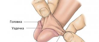
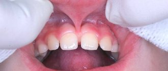
The frenulum of the upper lip in its physiological position at a distance of about 5-8 mm from the neck of the anterior incisors is woven into the gum. Sometimes the frenulum may be short. In this case, it is attached lower, at the diastema between the front incisors. In some cases, the attachment point is not visible at all. Diagnosing such a pathology is not difficult; it is visible during a routine visual examination. Since a short frenulum can lead to the development of various disorders, it is recommended to undergo plastic surgery.
Indications for plastic surgery
A short frenulum leads to impaired articulation; in complex cases, the mobility and functioning of the lips is reduced. Pathology can cause serious problems:
- In newborns, the sucking function is impaired, which interferes with normal feeding of the baby.
- Normal speech formation is disrupted, as there are difficulties opening the mouth and pronouncing labial sounds.
- Over time, malocclusion may develop and chewing functions may be impaired.
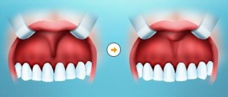
- When the frenulum is attached near the edge of the alveolar process, the interdental papillae are retracted and a diastema is formed.
- In the place where the gums are retracted, a deep gum pocket is created, in which dental plaque and tartar accumulate. In difficult cases, the roots of the teeth are exposed and an inflammatory process develops.
Since plastic surgery of the upper lip frenulum is a surgical intervention, before performing the operation the doctor assesses the condition and degree of deviation. Surgery is required in the following cases:
- A diastema has formed.
- The short frenulum was discovered during preparation for the installation of braces.
- There is gum recession and there is a risk of developing periodontal disease.
A neonatologist or speech therapist may also recommend plastic surgery if the disorder is detected at an early age. Plastic surgery is usually performed on children over 5 years of age, but if the sucking function is seriously impaired, the operation can also be performed on infants. Five years of age is explained simply. At this time, the loss of baby teeth and the formation of a bite begins.
Female reproductive system
October 23, 2015
The female genital organs are divided into internal and external. The female internal genitalia include the ovaries, fallopian tubes, cervix, uterus, and vaginal ligaments.
The ovaries (ovaries), or female gonads, are a paired gland of external and internal secretion of an oval shape, weighing from 5 to 8 g. It is located near the wall of the pelvis on both sides of the uterus. The ovaries perform two functions: they produce hormones (the most important of them are estradiol and progesterone) and produce eggs. Hormonal function is an important link in the body's hormonal system, regulating not only the function of the genital organs, but also the normal functioning of the female body as a whole. Despite age-related changes in the ovaries, their metabolic and hormonal function remains unchanged throughout life. Even before a girl is born, the development of future eggs begins in her developing ovaries. At approximately 5-6 months of pregnancy, the fetal ovaries contain 6-7 million future eggs, most of which are atretic before the birth of the girl. A newborn's ovaries contain approximately 400,000 immature eggs; subsequently no new eggs are formed. In childhood, atresia continues, and the number of eggs decreases even more. The immature eggs are surrounded by a thin layer of cells that form the follicle. At the onset of puberty, girls begin menstruation; In each menstrual cycle, several eggs mature, during which these cells divide twice, and the amount of genetic material they contain is halved. Through this process, called meiosis, each immature egg divides into four cells, of which only one forms a mature, fertilizable egg (ovum). A mature egg reaches 0.135 mm in diameter and is surrounded by a shiny membrane - zona pellucida. The human egg is very small, smaller than the period at the end of this sentence. The functions of three other cells, called polar bodies, are unclear; it is known that they will eventually degenerate.
The ovary is divided into zones of cortex and medulla. The cortex contains a huge number of follicles at various stages of maturation and also produces hormones. Primary follicles here transform into mature vesicular ovarian follicles, or Graafian vesicles. The latter contain the egg. After the surface of the ovary breaks through and the egg leaves it, a corpus luteum develops in place of the follicle, which subsequently, if pregnancy does not occur, atrophies and turns into a whitish body. The medulla contains connective tissue with blood vessels and nerves.
The fallopian tubes are a paired organ located on both sides of the uterus, extending from it to the side wall of the pelvis in the upper part of the high fold of the peritoneum (broad ligament of the uterus). The length of the pipe is 10-12 cm. It has narrow and wide ends. The narrow fallopian tube opens into the uterine cavity, and the wide one, like an octopus, covers the ovary. The wide part of the fallopian tube (funnel) has a large number of fimbriae. All this contributes to the fact that the egg released from the ovary enters the fallopian tube and moves along it to the uterus, which depends on the oscillatory movements of the cilia, which are equipped with the cells of the mucous membrane of the tube. Fertilization occurs already in the fallopian tube; later, the fertilized egg reaches the uterus and is fixed on its mucous membrane.
The cervix (cervix) is the lower part of the uterus that protrudes into the vagina. From the vaginal side, the cervix of a nulliparous woman looks like a smooth pink button with a rounded surface and a small hole in the center. Sperm penetrate the uterus through the cervical os; Through it, menstrual blood is released from the uterus. The cervical canal (the thin tube connecting the cervical os to the uterine cavity) contains numerous glands that produce mucus. The consistency of this mucus depends on the hormonal background and therefore changes at different stages of the menstrual cycle: immediately before ovulation or during the latter (when the egg leaves the ovary), the mucus becomes thin and watery; at other times it is thick and forms a plug that blocks the entrance to the cervix. There are no superficial nerve endings in the cervix, and therefore touching it causes almost no sexual sensations; Surgical removal of the cervix does not reduce a woman's sexual reactivity.
The uterus (uterus) is a hollow muscular organ intended for the intrauterine development of the fetus. Located in the pelvic cavity between the rectum and bladder. It is small in size and shaped like a pear. Its length is approximately 7.5 cm and its width is 5 cm. Its wide part (bottom) faces up and forward, and its narrow part (neck) faces down and back. Normally, the longitudinal axis of the uterus is oriented along the axis of the pelvis. When tilted forward, the body of the uterus forms an acute angle with the longitudinal axis of the vagina.
In this position, the uterus is held by wide uterine ligaments and folds of the peritoneum running from the edges of the organ to the side walls of the pelvis. The uterine cavity has the shape of a triangle. The fallopian tubes open in the upper corners, the lower corner passes into the cervical canal, connecting the uterine cavity with the vaginal cavity.
Like all hollow organs, the uterus has a three-layer wall. Its mucous membrane (endometrium) undergoes cyclic changes and is partially rejected during menstruation, which is accompanied by bleeding, and at the beginning of pregnancy a fertilized egg is implanted into it.
The muscular lining of the uterus (myometrium) is complex, consisting of three layers with different directions of muscle fibers. Outside, most of the surface of the uterus is covered with peritoneum. Where the uterus does not have a peritoneal covering, it is adjacent to connective tissue containing blood vessels, nerve fibers, and in places there are bundles of smooth muscle cells. This peri-uterine tissue (parametrium), extending in different directions to the walls of the pelvis, also provides fixation of the uterus.
The muscle wall is actively involved in labor and delivery.
Both functions of the uterus are regulated by hormones and chemicals that also cause the uterus to enlarge during pregnancy.
On both sides of the uterus there are fallopian tubes and ovaries attached to it by lateral ligaments. It is necessary to name another ligament, the uterosacral ligament, which, starting from the posterior surface of the cervix, goes around the rectum on both sides and is attached to the periosteum of the sacrum. Inside this ligament are bundles of nerve fibers extending from the spinal cord along which sexual impulses rush to the uterus, vagina, vulva and clitoris.
During pregnancy, the uterus gradually increases in size, and its muscle mass increases many times over. The pregnant uterus occupies a significant part of the abdominal cavity. At the time of birth, the muscles of the uterus contract spasmodically, expelling the fetus.
In cases where the uterus is rigidly fixed by adhesions that occur after operations or as a result of an inflammatory process, a woman may feel pain during sexual intercourse; this situation requires surgical intervention.
The vagina is a hollow muscular cylinder. Its internal expanded end covers the cervix, and the external one passes into the genital slit. The inside of the vagina is lined with a mucous membrane with a large number of folds. On the anterior and posterior walls of the vagina, these folds become higher, forming longitudinally oriented columns of folds. The anterior column of folds, located on the anterior wall of the vagina, is better expressed than on the posterior wall. Below it is a longitudinally oriented protrusion, the urethral carina of the vagina. It should be noted that the anterior wall of the vagina in the upper third is adjacent to the bottom of the bladder, and in the remaining area it is fused with the wall of the urethra. On the outside of the vagina there is a layer of loose fatty tissue, as a result of which it can easily expand and shift. Despite the fact that there are no glands in the vaginal mucosa, it is always moist due to the secretion of lymph from the tissue and mucus from the cervix. Normally, vaginal mucus has a whitish-milky color and its reaction is acidic. The acidic environment of vaginal contents prevents the development of pathogenic microorganisms. Under normal conditions, the amount of vaginal discharge is insignificant, and it is not released outside. The appearance of vaginal discharge of an unusual color and odor usually indicates a painful condition.
The walls of the vagina contain a large number of nerve endings, the irritation of which enhances sexual sensations. During sexual arousal, the walls of the vagina, due to the rush of blood, swell, making it narrower and more sensitive.
In women who have not given birth, the entrance to the vagina can be very narrow (slit-like) and almost without a lumen, but in women who have given birth, it is open and is an oval-shaped hole (after partial deformation of the perineum during childbirth). In girls, the vaginal opening is almost half covered by the hymen. The vagina, like an inflatable balloon, can change its shape and size. It can expand, creating conditions for the passage of the baby’s head during childbirth, or it can shrink so much that it covers the finger inserted into it on all sides. Despite its ability to contract, a woman's vagina cannot enclose the penis during intercourse so tightly that physical separation becomes impossible. The mating that sometimes occurs in dogs is mainly due to the expansion of the bulbar part of the penis.
The inner lining of the vagina is similar to the oral mucosa. The vaginal mucosa provides hydration. There are no secretory glands in the vagina, but it is rich in blood vessels. The endings of sensory nerve fibers are present at the entrance to the vagina, and in other parts of it there are relatively few of them. As a result, the deeper part of the vagina (about two-thirds) is relatively less sensitive to touch or pain.
The external female genitalia include the mons pubis, labia majora, labia minora, clitoris, hymen, vaginal vestibule, and perineum.
Pubis (mons veneris) is an elevation arched above the entrance to the vagina, the width of a thumb. The pubis consists of fatty tissue covered with skin and hair. There are many nerve endings in this area, so touching and/or applying pressure to it can cause sexual arousal.
The labia majora (labia majora) are rounded folds of skin that border the genital opening on the sides. Their length is 6-8 cm, thickness 2-3 cm. They connect in the pubic area. In the thickness of the labia majora on both sides there are large glands of the vestibule (Bartholin's), similar to the bulbourethral glands in men, producing mucus that moisturizes and alkalizes the acidic contents of the vagina during sexual arousal. In addition, numerous sebaceous and sweat glands are scattered in them.
When the thighs are closed, the inner surfaces of the labia majora touch and often completely cover the genital slit, although in some cases they do not cover the protruding labia minora and clitoris.
The labia minora (labia minora) are located inside the labia majora, parallel to them and usually almost 2 times smaller in size. The space between the labia minora is called the vestibule of the vagina. At the top, the labia minora meet to form a fold around the clitoris. This fold is called the foreskin of the clitoris. The thickness of the labia minora contains a dense venous network and Bartholin glands; each of them has a small duct that opens on the inner surface of the lip, near the vestibule of the vagina.
The clitoris is located at the upper ends of the labia minora and is analogous to the penis. Its length is 1-1.5 cm. It consists of 2 cavernous bodies, which are attached by their ligaments (legs) to the lower branches of the pubic bones. In the head of the clitoris, as in the head of the penis, there is a large number of nerve endings that cause its high sensitivity and excitability. Below the clitoris is the external opening of the urethra, on both sides of which the paraurethral ducts (Skeene's ducts) open. Between the clitoris and the external opening of the urethra there is an unpaired corpus cavernosum, similar to the bulbous section of the male urethra.
The vestibule of the vagina (introitus vaginal) is a space bounded in front by the clitoris, behind by the posterior commissure of the labia majora and on the sides by the inner surface of the labia minora. The bottom of the vestibule is the hymen. In the vestibule of the vagina, the external opening of the urethra, the opening of the vagina (in the center of the hymen) and the large glands of the vestibule (Bartholin's) open. On each side of the vestibule there is a cavernous body, the bulb of the vestibule. The area that includes the inner surface of the labia minora, the vestibule of the vagina and the urethral carina of the vagina on its anterior wall is the most sensitive area of the vulva after the clitoris.
The hymen (hymen) is a thin membrane of mucous membrane that closes the entrance to the vagina in women who have not had sexual intercourse. The shape and size of the hymen varies.
The perineum is a collection of tissues located between the posterior commissure of the labia majora and the apex of the coccyx. The part from the posterior commissure to the anus is called the anterior perineum, and from the anus to the top of the coccyx the posterior perineum. In the thickness of the perineum are the fascia and muscles that make up the pelvic floor.
Contraindications
Plastic surgery of the frenulum of the upper lip is not a complex operation; however, there are a number of contraindications for which the operation cannot be performed. These include:
- Diseases of the oral mucosa with frequent relapses.
- Multiple caries. In this case, treatment is required first.
- Common infectious diseases in the acute stage.
- Blood diseases.
- Lesions of the nervous system, mental disorders.
- Oncological diseases.
Short frenulum of the tongue in a child
The lingual frenulum is a fold of the oral mucosa that connects the floor of the mouth to the undersurface of the tongue. The frenulum of the tongue performs a very important function - it fixes the tongue to the soft tissues of the oral cavity, preventing tongue retraction.
Normally, the frenulum stretches from the middle of the lower surface of the tongue, but in some cases the structure of the frenulum is disrupted, thereby shortening it, that is, the upper end of the frenulum is not in the middle, but at the tip of the tongue. Anomaly of the frenulum of the tongue requires observation and even treatment.
You can see a shortened frenulum of the tongue with the naked eye; the following signs indicate it:
- low mobility of the tongue (the child cannot lift the tongue or stick it out);
- bifurcation of the tip of the tongue (a short frenulum pulls the tip of the tongue);
- difficulty feeding in infancy (the baby cannot properly latch onto the mother's nipple or bottle).
Untimely treatment of a shortened frenulum can lead to many consequences, the main ones being:
- the formation of unclear speech, difficulties begin when pronouncing sounds;
- deformation of the lower teeth and lower jaw;
- spaces between teeth;
- malocclusion.
It is recommended to trim the frenulum of the tongue for a child from birth to one year. As a rule, the problem of a short frenulum of the tongue should be noticed in the maternity hospital during the examination of a newborn, when it is much easier to trim it than at an older age. But this does not always become noticeable to the surgeon immediately, so the problem appears already in preschool age, when the child begins to have a clear deviation in speech.
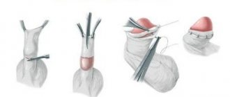
The operation to cut the short frenulum of the tongue should be performed by a dentist. There are several different types of surgery, depending on the age of the child and each specific case:
For babies.
The most suitable age for correcting a frenulum is from 0 to 9 months; it is during this period that the frenulum does not have nerve endings, which makes it easier to carry out the procedure without anesthesia. As a rule, babies undergo a frenotomy - they cut across the skin fold with scissors or a scalpel. In this case, no stitches are required.
For children from 9 months to 5-6 years.
After 9 months, the frenulum thickens, so surgery will require painkillers and possible sutures.
For school-age children and teenagers.
Surgery for older children is more complex as it requires local anesthesia and stitches. After a couple of weeks, the stitches are removed. The postoperative period requires some diet to restore the sutures.
Today, cutting the frenulum of the tongue with a laser has become popular; the procedure takes only one step and does not cause any discomfort. Only a specialist can assess which method of operation will be needed in each specific case.
Carrying out the operation
The operation does not require any serious preparation. If there are dental diseases, they are treated first. The doctor must sanitize the oral cavity to avoid the risk of developing an infectious process. Plastic surgery is done using different methods:
- Frenulotomy (dissection). This method is used to correct a narrow frenulum without a visible attachment point. The doctor cuts it transversely. After this, sutures are applied in the longitudinal direction.
- Frenectomy (excision). Unlike the previous method, frenectomy is performed to correct a short and wide frenulum. During the operation, the doctor tightens it and makes an incision along the ridge. The interdental papilla is also excised. If there is a large diastema, the tissue between the end parts of the incisors is also excised.
- Frenuloplasty. With this type of adjustment, the area where the bridle is attached moves. This operation is performed by two methods, which differ from each other in the way of forming the mobilized flap. To do this, the doctor trims the mucous membrane so that it can be moved along the periosteum. The operation with cutting and mobilization of the mucosa is more complex than the two previous types, so it is performed less frequently.
Regardless of the method chosen, the operation is performed under local anesthesia and takes about 15 minutes.
Laser plastic surgery
Laser plastic surgery is a relatively new, but already very popular method. The laser beam vaporizes the tissue of the frenulum, while simultaneously sealing the edges of the resulting wounds. To relieve pain, the removal site is treated with an anesthetic gel. An important advantage of laser operations is a short postoperative period and the absence of scars.
When using a laser, there is almost completely no risk of inflammation, infection and other complications. The laser beam disinfects the area of treatment, and thanks to the sealing of the edges of the wounds, the operation is carried out bloodlessly.
The rehabilitation period after plastic surgery is about 5 days. At this time, you must follow the doctor's recommendations so as not to injure the operated area.
If you have the symptoms described in this article, be sure to make an appointment at our clinic.
Don't self-medicate! Even the smallest problem, if not treated correctly, can significantly complicate your life.
By contacting us, you can be sure that:
- Get high-quality and free consultation .
- You will receive the best prices for treatment and the opportunity to receive a special promotional price.
- Only modern equipment and materials will be used.
- You will be treated by professional doctors with many years of experience.
- We offer treatment on credit or in installments. There is also the possibility of obtaining a tax deduction.
- We work seven days a week and without a lunch break, from 9 a.m. to 10 p.m.
+7 (495) 132-02-96
Make an appointment