Erosion of tooth enamel is a fairly common problem and, contrary to what many people believe, not harmless: in addition to the unpleasant cosmetic effect, the pathology negatively affects the condition of the teeth and requires immediate dental care.
In this article we will talk about the causes of erosion on teeth and the methods of solving the problem offered by modern dentistry.
Causes of erosion and consequences of pathology
Erosion of tooth enamel is a type of non-carious lesion. The first signs of the disease are the appearance of small, slightly protruding areas on the surface of the teeth; the course at the initial stage is without obvious symptoms. However, over time, the enamel’s sensitivity to temperature factors increases, the spots gradually darken, unpleasant and even painful sensations appear, and hard tissues begin to wear out very quickly.
Important!
At the moment, the reasons for the development of pathology are not precisely known!
However, experts name a number of factors that can provoke the appearance of erosion. Among them:
- disturbances in the functioning of the endocrine system;
- causing mechanical damage (including the use of home methods for whitening tooth enamel, hygiene with a hard brush, abuse (constant use) of dental veneers with abrasive particles, etc.)
- exposure to certain chemicals (eg, vinegar, sour drinks, citrus fruits);
- uneven chewing load (standard reasons - malocclusion, loss of a unit of dentition without timely restoration);
- constant interaction with chemicals, contact with mineral or metal dust (usually associated with professional activities).
- long-term use of potent pharmaceutical drugs.
By delaying contacting the dentist, you risk waiting until the damaged top layer leads to dentin damage.
Medical Internet conferences
Introduction. Currently, dental erosion occupies a significant place among diseases of hard dental tissues. There are many opinions about the origin of erosive defects, and this issue has not been fully studied. This topic gives rise to a lot of controversy and questions among scientists and doctors, and therefore requires more attention. Research results indicate a significant increase in the prevalence of dental erosion in the last 10 years. Thus, when examining a population group, 47.2% of people with dental erosion were identified, while 10-15 years ago there were no more than 5-7% of such patients. When analyzing the frequency of non-carious dental lesions, based on patients’ visits to the dental clinic, 29.5% of people with dental erosion were identified. Meanwhile, 10-15 years ago there were only 24 such patients. Moreover, the disease was observed mainly in women (84.9%) aged 25-30 years. The combination of erosions with hormonal disorders (including dysfunction of the thyroid and gonads) accounted for more than 75% of cases.
Objective: to analyze literature data on the etiology of erosive defects and their impact on the quality of life of patients.
Tasks:
1) characterize the hypotheses of the origin of erosions of hard dental tissues
2) to study the mechanism of occurrence of erosions of enamel and dentin
3) assess the quality of life of patients with erosive dental defects
4) create a draft treatment plan for patients suffering from erosive dental changes.
Materials and methods: scientific articles and works, domestic and foreign scientific literature on dentistry were analyzed, clinical cases of erosion of hard dental tissues of varying severity of the pathological process were analyzed.
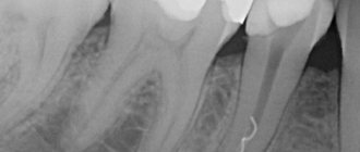
Results and discussions. Hard tissue erosion (erosion) (from the Latin erosio - “corrosion”) is the progressive loss of tooth enamel and dentin. In foreign literature, both narrow terms are used: “attrition”, “abrasion”, “erosion”, “abfraction”, and broader ones: “toothwear” and “tooth surface loss”. In European literature, the most common point of view is that erosion is a more important factor in the loss of hard dental tissues than abrasion due to contact of tooth surfaces. Occurs after teething. The affected area can be located on the vestibular and palatal surfaces and has a round, cup-shaped shape with dense, smooth, flat edges. This is a differential diagnostic feature when making a diagnosis. The upper incisors are primarily affected, less commonly the canines and premolars. It is extremely rare that erosions occur on the teeth of the lower jaw. Probing and percussion are painless. EDI 2-4 µA. The oral mucosa is without visible pathological changes.
2. The causes of erosion have not been clearly established.
According to ICD-10, the pathological condition of hard dental tissues is divided into two large groups:
· “Disorders of development and teething”
· “Other diseases of dental hard tissues.” K03
Tooth erosion refers to diseases of the hard tissues of teeth. K03.2
K03.2 Tooth erosion:
K03.20 Professional;
K03.21 Caused by persistent regurgitation or vomiting;
K03.22 Due to diet;
K03.23 Caused by drugs and medications;
K03.24 Idiopathic;
K03.28 Other specified dental erosion;
K03.29 Dental erosion, unspecified.
The first four causal factors of this classification reflect the chemical theory of the development of dental erosion present in the medical literature of previous years, which in turn considers the impact of aggressive chemical agents on the enamel as the leading causes:
I. External factors:
1) type of diet: consumption of acid-containing foods and drinks (marinades, pickles, citrus fruits, fruit and berry juices, sweet carbonated drinks, etc.)
2)work in hazardous industries associated with inhalation of acid fumes, metal and mineral dust particles
3) The effect of a number of medications on tooth enamel, for example, acid (acetylsalicylic and ascorbic), gastric juice preparations, hydrochloric acid.
II. Internal factors:
4) Dental erosion can be caused by chemically aggressive contents of the stomach and duodenum with a low pH content in chronic gastroesophageal regurgitation that occurs with gastroesophageal reflux disease, as well as with combined duodeno-gastroesophageal reflux disease. Erosive lesions are observed in individuals with hiatal and diaphragmatic hernias and those suffering from bulimia.
Damage from internal factors usually occurs on the palatal surfaces, and from external factors - on the buccal surfaces.
D. A. Entin saw the cause of erosion in neurodystrophic processes that cause decalcification of hard tooth tissues. However, no one can explain why erosions occur in some cases and wedge-shaped defects in others. Their occurrence may be associated with a violation of mineral metabolism due to endocrine or other disorders in the body and, accordingly, in the dental pulp. This is confirmed by the results of clinical observations and data from radioimmunological studies, which indicate the presence of clear preceding and concomitant dysfunctions of the thyroid gland in patients with erosions of dental enamel. Thus, Yu. M. Maksimovsky et al., analyzing the causes of erosions, assign an important role to endocrine disorders and, above all, hyperfunction of the thyroid gland. It was noted that dental erosions in patients with thyrotoxicosis were detected 2 times more often than in persons with normal thyroid function; a direct connection was established between the intensity of dental damage and the duration of thyrotoxicosis. As the duration of the disease increases by 1 year, the number of patients with erosion of hard dental tissues increases by 20%.
Dr. Kim McFarland, a dental surgeon and professor at the College of Dentistry at the University of Nebraska Medical School in Lincoln, USA, notes an increase in the number of patients with erosion of tooth enamel over the past 25 years, which is associated with uncontrolled consumption of carbonated sugary drinks.
A number of researchers (Baume, Port and Eidler) associate tooth erosion with excessive mechanical stress on the enamel, namely the use of hard toothbrushes, whitening toothpastes and powders with increased abrasiveness, as well as improper brushing techniques - a predominance of horizontal movements.
The combination of several predisposing factors accelerates the course of erosion and aggravates its severity. For example, drinking large amounts of very low pH drinks causes tooth surface loss when combined with brushing immediately after an acid attack on the teeth.
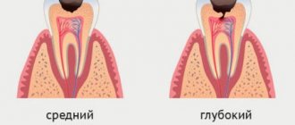
Yu. M. Maksimovsky details the clinical manifestations of erosions and distinguishes three degrees of damage, based on the depth of the hard tissue defect:
I degree (superficial, initial) – with damage to only the upper layer of enamel
II degree (medium) – with damage to the enamel throughout the entire depth up to the enamel-dentin border.
III degree (deep) – with damage to the entire enamel and the upper layer of dentin.
E.V. Borovsky et al., distinguish two stages of damage: initial (enamel erosion) and severe (enamel and dentin erosion).
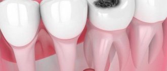
In degrees 1 and 2, the lesion is white with a shiny surface; in degrees 3, brown or light yellow pigmentation appears.
Dental erosion is usually characterized by a chronic course, but there are two clinical stages of erosion: active and stabilized.
The active stage is characterized by a progressive course and loss of tooth tissue, accompanied by hyperesthesia and the disappearance of the shine of the erosion surface. In the active phase, changes in the size of erosion occur every 1.5-2 months.
The stabilized form of erosion of hard tooth tissues is characterized by a calmer, slower course, and the shiny surface of the enamel in the affected area is preserved. There is no change in its size for 9-11 months. A transition from a stabilized form of erosion to an active one is possible, especially if the background pathology worsens.
3. Pathogenesis
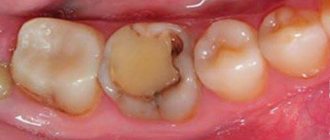
Unlike dental caries, where there is superficial, subsurface demineralization of the enamel, during erosion, superficial foci of demineralization are formed, which gradually cover the tooth enamel layer by layer. The microhardness of the enamel in the area of erosion is significantly reduced, and foci of demineralization of the enamel surface are noted. When studying the ultrastructure of enamel during tooth erosion using a scanogram, it was noted that the enamel in the area of erosion and in adjacent areas is characterized by a reduced degree of mineralization and the presence of destructive changes: in some areas, enamel prisms are clearly visible, interprismatic spaces are pronounced, and in others, enamel prisms and interprismatic spaces indistinguishable due to demineralization. Hydroxyapatite crystals of various shapes. In areas adjacent to erosion, they do not have clear boundaries or have a regular shape, but are large. Crystals of enamel hydroxyapatite with varying densities are visible on the surface of the enamel, indicating uneven mineralization. There are also distinct changes in dentin during tooth erosion: areas with a dense arrangement of crystals are observed. Dentinal tubules can be obliterated or non-obliterated. The structure of the substance that obliterates the dentinal tubules is specific and close to that during abrasion, however, along with the indicated areas of demineralization, accumulations of bacteria were found that mask the contours of the enamel prisms.
Comparative electron microscopy (SEM) of the central erosion zone also showed the presence of significant structural changes in both the superficial and deeper layers of damaged dental tissue. The active stage of the process is characterized by the loss of both enamel substance and dentin in large areas that have undergone destructive changes.
In the cervical region of teeth with erosive defects, an intermittent but quite clearly visible boundary between the crown and root is visible. In all studied cases, the crown enamel was layered on the root cement.
4.Quality of life of patients with dental erosion.
Enamel is the protective shell of the tooth. The process of enamel erosion is irreversible and creates problems for a person for life.
Patients complain of an aesthetic defect, the presence of a defect in the cervical area, tooth sensitivity, and tissue loss.
According to our data (based on the number of visits to the clinic), about 15% of patients are aged 16-42 years.
We observed 3 patients with varying degrees of dental erosion.
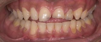
1) Patient A., 23 years old; preliminary diagnosis: dental erosion, stabilized form; mild severity according to ICD-10 K03.2. Complaints about dissatisfaction with the color of teeth. Objectively: on the vestibular surface in the cervical area of 1.1 and 2.1 teeth there is a defect affecting only the upper layer of enamel, the oral cavity is sanitized, the oral mucosa is without pathological changes, IG - 1.7 (satisfactory).
History: consumption of freshly squeezed citrus juices, professional cleaning with Air-flow 2 times, tried to use whitening toothpastes.
2) Patient K., 28 years old; preliminary diagnosis: active stage of dental erosion, moderate to severe degree according to ICD-10 K03.22
Complaints about tooth sensitivity, aesthetic defects, yellow teeth.
Objectively: on the vestibular surface in the cervical region, 1.1 teeth have a pronounced defect within the enamel and 2.1 teeth have affected the entire enamel and the upper layer of dentin. The oral cavity is sanitized, the oral mucosa is without pathological changes IG-2.2 (unsatisfactory).
History: consumption of citrus fruits, incorrect selection of oral hygiene products and items, incorrect method of brushing teeth with a predominance of horizontal movements.
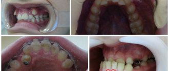
3) Patient L., 42 years old; preliminary diagnosis: active form of dental erosion, severe degree according to ICD-10 K03.21
Complaints about the presence of defects in the cervical area, tooth sensitivity and aesthetic defects.
Objectively: erosive defects in the frontal group of teeth of the upper and lower jaw, in the cervical area there are lesions of brown pigmentation. The oral cavity is sanitized, the oral mucosa is without pathological changes, IG-1.6 (satisfactory)
History: occupational hazards, chronic gastroesophageal regurgitation, traumatic occlusion, crowded teeth.
Thus, regardless of the severity of erosion, the quality of life of these patients suffers to one degree or another. Even if practically nothing bothers the patient at first, in the future, in the absence of correction of etiological factors and specialist interventions, the symptoms increase like an avalanche (according to patients with more pronounced defects), which forced us to try to create an algorithm for treatment and preventive measures (draft treatment plan) for this group patients.
5. RECOMMENDATIONS
Enamel erosion is not just an external problem, but a serious disease, and therefore the attitude towards treatment is no less serious.
- A thorough ascertainment of the patient’s history and current condition with the involvement of related specialists (therapist, gastroenterologist, endocrinologist, pediatrician, etc.), gastronomic preferences, and features of professional activity will significantly identify the possibility of correcting patient-dependent factors or, at a minimum, recording them in the outpatient dental record. sick.
- Treatment of patients with erosion should be comprehensive and long-term.
- Strict diet (except citrus fruits, berries, sweets, carbonated drinks, fresh juices containing vitamin C, canned foods). Include protein in your diet to strengthen the protein matrix of enamel and collagen fibers.
- Select products (pastes containing organic calcium, with hydroxyapatite) and hygiene items (correction of the rigidity and structure of the brush bristles, excluding the use of toothpicks), as well as teach the correct method of brushing teeth (vertical movements).
- Remineralizing therapy (Rocs medical minerals gel, Remars gel, Clinpro™ White Varnish,) Tooth Mousse, “Belagel Sa/R” “VladMiVa”) in a clinical setting in the form of applications and mouth guards (preferably individually). At home daily, perhaps constantly, but necessarily regularly, depending on the degree - office-based in combination with fluoride applications to prevent concomitant caries and to strengthen the crystal lattice of hydroxyapatite.
- Avoid ultrasonic teeth cleaning, home and professional whitening, and Air-Flow teeth cleaning.
- For professional hygiene, use pastes with minimal abrasiveness “fine”.
- Restoration if necessary, after complex treatment. It is possible to use the Icon technique, as well as the use of a desensitizer (SHIELD FORCE PLUS).
- clinical examination with photographic recording of the result.
Conclusions:
1. In the etiology of erosion of hard dental tissues, the following factors interact: exogenous (occupational hazards, dietary habits) and endogenous factors (metabolic disorders, endocrinopathies, bruxism, diseases of the gastrointestinal tract) in combination with improper oral care.
2.The main mechanism is demineralization of the enamel.
3. Regardless of the severity of erosion, the quality of life of these patients suffers to one degree or another. Even if at first there is practically nothing bothering the patient, in the future, in the absence of correction of etiological factors and specialist interventions, the symptoms increase like an avalanche.
4. The algorithm of treatment and preventive measures for this group of patients should include:
-correction of both external and internal etiological factors;
-specialized treatment by a dentist before the required restoration with the use of remineralizing and fluoride-containing drugs up to restoration (using filling materials from the group of compomers or GIC);
- general treatment with mandatory medical examination for a long time (lifelong).
Why you can't ignore the problem
If the problem is not recognized in time and treatment is not started, very serious complications are possible, namely:
- distribution of dark spots over the entire surface of the tooth;
- violation of the uniformity of enamel color (up to the transparency of the cutting edge);
- increased abrasion of enamel, accelerated wear of teeth;
- increased sensitivity to temperature influences, the appearance of painful sensations.
When the lesion spreads to the dentin, rapid destruction of the hard tissues of the tooth occurs, as a result - the development of various dental diseases
The widest risk group by age is people of middle age, but the pathology is often found in children.
Diagnosis of non-carious lesions
The main signs of destruction of dental tissues:
- change in enamel color;
- change in the structure of the tooth surface, which becomes wavy, dotted or grooved;
- visual reduction of the coronal part, formation of round defects with a diameter of up to 5 mm.
Also, one of the main symptoms is increased sensitivity of the enamel (hyperesthesia). In the first stages, a reaction to hot and cold food appears; over time, pain arises from chemical irritants (sour, sweet, salty) and the slightest tactile influence.
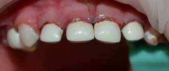
Erosion on baby teeth
In children, the most common cause of the development of non-carious lesions is excessive consumption of liquids high in sugar and acids (milk and juice). Therefore, if the baby refuses to fall asleep without a bottle, it is better to pour regular water into it. It is also important to take care of oral hygiene: regularly cleaning them from bacterial plaque will help prevent the development of erosion on baby teeth.
Other problems that lead to erosion in children are crowded teeth and malocclusions.
Carefully monitor the condition of the tooth enamel in children, contact your dentist immediately when the first signs of the disease appear!
Non-carious lesions of teeth in children
Non-carious lesions in children most often belong to group I, that is, tooth germs are destroyed or completely die at the development stage (the period of intrauterine life of the child).
This may occur due to genetic disorders, toxic effects of medications taken by the expectant mother, as well as intrauterine infections. Excess or deficiency of vitamin D, as well as excessive fluoride concentrations, also have a detrimental effect.
Early toxicosis of a woman during pregnancy can also disrupt the mineralization of a child’s teeth.
Diagnosis of erosion treatment
To make a diagnosis in the early stages, differential diagnosis is necessary, taking into account various manifestations - from increased tooth sensitivity to problems with the pronunciation of whistling sounds. If stains on the teeth have already appeared, a visual inspection is sufficient.
The clinical picture of erosion of tooth enamel is similar to the manifestations of superficial caries (the disease is also characterized by a rough surface of the teeth) and erosion with a wedge-shaped defect (characterized by lesions along the neck of the tooth). Only a dentist can make a correct diagnosis!
After diagnosing erosion, treatment is prescribed, which usually includes a number of procedures:
- calcium electrophoresis (restoration of the mineral composition of enamel, saturation of tooth tissue with useful microelements and minerals);
- carrying out remineralization (2-3 week course of applications with F and Ca followed by application of fluorinated varnish to the enamel);
- restorative therapy (vitamin complexes, drugs with a high content of F and Ca).
Elimination of aesthetic effects is ensured by professional cleaning of the enamel with bleaching and protection of the tooth surface with fluoride varnishes and gels.
In case of serious damage to the enamel, they resort to dental prosthetics with veneers (ultra-thin overlays fixed on the outside of the teeth perfectly hide the defects); the most advanced cases may require crowns.
Do not get carried away with advice on combating erosion using traditional medicine: self-medication in this case is dangerous, the thinning of the enamel cannot be stopped by such means, and it’s easy to aggravate the situation!
The time and complexity of treatment directly depends on how deeply the erosive processes have spread. Proper hygiene and regular preventive examinations will allow you to avoid long-term exhausting therapy and expensive prosthetics.
The best treatment is prevention
To avoid the problem, follow these simple recommendations:
- do not forget to brush your teeth twice a day, but do not do it immediately after eating acid-containing food (delay the procedure for 30-60 minutes);
- choose toothbrushes with natural bristles, soft or medium hard;
- make an informed choice of toothpaste: avoid constantly using whitening pastes containing abrasive particles; give preference to fluoride-containing pastes that strengthen and mineralize the enamel;
- leave foods with neutralizing properties (a piece of hard cheese, a glass of milk, etc.) at the end of the meal;
- Reduce the amount of foods containing aggressive acids in your diet.
A responsible approach to maintaining oral and dental health is a guarantee of reliable protection against dental problems!