Home / Cancer Treatment / Treatment of Oral Tumors
- Doctors
- Tomotherapy
- Reviews
- Cost of treatment
- Risk factors
- Symptoms
- Staging
- Diagnosis and treatment
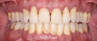
Various malignant neoplasms develop in the oral cavity, and in most cases it is squamous cell carcinoma. Based on location, tongue cancer is most often recorded than others. Its anterior two-thirds account for 75% of malignant lesions. The next most common localizations are malignant neoplasms of the mucous membrane of the cheeks and the floor of the mouth.
In men, cancer of the oral mucosa is three times more common than in women, with the peak incidence occurring in old age, although the pathology occurs in all age groups, including children.
Tomotherapy for oral cancer
The use of external beam radiation therapy is accompanied by two problems: damage to healthy tissue and insufficient regression of the tumor. Side effects sometimes completely neutralize the achieved treatment result and increase the risk of developing postoperative complications. Intensity modulated radiation therapy (IMRT), and in particular TomoTherapy HD technology, can solve these problems and increase the effectiveness of radiological treatment.
TomoTherapy HD is a complex that combines a computed tomograph and a modern particle accelerator. The system allows you to accurately deliver a dose of radiation to a tumor, no matter how complex it is (localize the tumor, plan treatment and carry it out), protecting healthy tissue from radiation exposure. This approach can significantly reduce the risk of unwanted manifestations.
The ability to deliver a dose of radiation with minimal impact on healthy tissue allows for a more pronounced effect on the tumor and reduces the likelihood of severe side effects in the future
Reviews
I would like to express my deep gratitude to the entire team of the radiation therapy center - attending physician Maria Viktorovna Aglullina, nurse Gulfiya Khafizova, medical registrar Venera Diyarova - for their kind, sensitive attitude towards me. During this difficult period of struggle and treatment, the attitude of the people around you and the medical staff is very important. I wish all employees good health, happiness and love! Thank you so much for everything!
Patient from Kazan
July 30, 2020
All reviews
Rushana's story, lung cancer
“Hello, my name is Rushana, I live in the city of Kazan and I ended up in this center, like many people, probably unexpectedly. Because for most of our lives we do not imagine that we might encounter anything else, that there might be some serious difficulties in our lives that we have not yet overcome.
All stories
Initial or “zero” stage
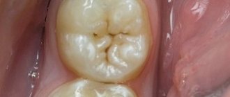
If you are trying to understand what gum cancer looks like and its initial stage, then in vain. You will not find photos on the Internet with characteristic visual manifestations of the disease, because there are no external signs as such. The only thing that can be alarming is the very first symptoms, namely, slight redness and swelling of the gums, its hardening, which persist for a long time. In this case, the affected area usually has a clear localization and is limited from other tissues; it seems to rise above them.
The disease is asymptomatic. However, dentists will be able to suspect its presence and diagnose it if you undergo annual preventive examinations, and this will significantly increase the chances of successful treatment.
Cost of treatment for oral tumor
| Name of service | price, rub. | Unit measurements |
| Consultation with an oncologist and radiotherapist | 1 500 | PC. |
| Consultation with a pediatric oncologist | 0 | PC. |
| Repeated consultation with specialists | 500 | PC. |
| Primary topometry on a specialized computed tomograph | 15 000 | procedure |
| Repeated topometry on a specialized computed tomograph | 7 000 | procedure |
| Primary dosimetric planning of radiation therapy (tomotherapy) | 20 000 | PC. |
| Repeated dosimetric planning of radiation therapy (tomotherapy) | 7 000 | PC. |
| Radiation therapy (tomotherapy), including IMGRT (*) | 315 000 | well |
| Radiation therapy (tomotherapy) stereotactic radiofrequency surgery (*) | 315 000 | well |
| Accompanying drug therapy: intravenous administration in the treatment room (excluding the cost of medications) | 1 000 | procedure |
| Accompanying drug therapy: intramuscular administration in the treatment room (excluding the cost of medications) | 200 | procedure |
| Topometric marking | 750 | procedure |
The type of radiation therapy and the number of sessions of the course are determined by the medical commission individually for each patient based on the location, nosology of the tumor and taking into account the medical history.
Free online consultation
Sign up
Risk factors
Most malignant tumors of the oral cavity arise in already changed tissues. Usually these are long-term inflammatory changes and various precancerous conditions. Precancer is a special condition of tissues in which at some point their malignant degeneration may occur, although a benign tumor and a complete return to normal state may be a possible outcome. The causes of precancerous conditions and their progression into a malignant process are not known with certainty. Scientists suggest that this depends on the immunobiological status of a person and the characteristics of exposure to the carcinogen.
Factors that contribute to the appearance of precancerous changes can be both external damaging agents and various disorders in the body:
- Constant mechanical impact - malocclusion, poorly fitting dentures.
- Chemical agents - alcohol, tobacco smoke, spices, industrial irritants.
- Temperature agents - constant consumption of very hot food, hot tobacco smoke, work in hot shops.
- Biological agents: various microorganisms of the oral cavity, both frankly pathogenic and opportunistic, which make themselves known only under favorable conditions.
- Ionizing radiation, including due to medical procedures.
- Diseases of the digestive system.
- Some systemic lesions, in particular lupus erythematosus.
Among these factors, one of the most important is biological. There has been a proven connection between precancerous changes in the oral mucosa with caries and gum disease due to excessive bacterial growth. The second, but no less important factor is smoking - the disease is registered in smokers 6 times more often than in non-smokers.
Symptoms of gum cancer
Gum tumors are divided into:
- exophytic - growing outward and forming a node;
- endophytic - spreading deep into the gums, in the form of a flat infiltrate or ulcer;
- mixed - a combination of two growth options; as a rule, this is a far advanced or recurrent process.
I detect a knot on the surface of the gum at an early stage, because it gets in the way, the tongue “feels” for it, and it gets injured when brushing your teeth or while eating. Infiltrates grow faster and are found later, when complaints of pain or bleeding appear.
Cancer in the initial stage does not hurt, especially since the oral mucosa is accustomed to irritating, spicy foods and hot foods. Temporary discomfort while eating does not cause everyone anxiety with a desire to see what is happening there. Gum cancer can masquerade as toothache, forcing you to see a dentist. Most often, it is the dentist who discovers the malignant process.
In stages 3-4, when cancer grows into the jaw, destroying the bone, the pain becomes constant, sharply intensifying while eating, and due to basic malnutrition leading to significant loss of body weight.
A large cancerous ulcer is accompanied by inflammation of the surrounding tissues and decay, causing intense pain radiating to the ear and temple. Secondary infection of the cancerous infiltrate is inevitable; it is manifested by a “heavy” putrid odor. The patient cannot open his mouth due to a painful spasm of the masticatory muscles - trismus. The general condition worsens, the temperature rises, and weakness increases.
Citizens who abuse alcohol drown out the pain by increasing the dose of alcohol, and complain of the appearance of a painful and often inflamed metastatic lymph node under the jaw or on the neck. The presence of various microflora in the mouth complicates metastatic conglomerates in the lymph nodes with secondary infection; when the size is more than 3-4 cm, a zone of decay is formed in the center of the lymph node, often with the formation of a fistula. During this period, symptoms of intoxication with high fever appear.
A reflex increase in salivation with impaired swallowing is characteristic. Salivation is very profuse, so it is possible that saliva may be thrown into the respiratory tract at night with the development of aspiration pneumonia. All malignant tumors lead to increased thrombosis and ease of development of bacterial infection in a weakened body. An additional negative factor is malnutrition up to complete starvation with exhaustion and the development of the fatal anorexia-cachexia syndrome.
Distant metastases are not typical for gingival carcinoma, but recurrences are common after treatment. Cancerous destruction of oral tissues with frequent bleeding from vessels destroyed by the tumor against the background of chronic inflammation in metastatic lymph nodes leads to a serious condition and death.
Clinical manifestations
Symptoms of oral cancer may include:
- compaction and swelling of the affected area;
- long-term non-healing ulcer;
- white or red spots on the mucous membrane;
- weakening of tooth roots;
- bleeding gums;
- speech disorder;
- unexplained weight loss;
- soreness in the mouth;
- general malaise.
Most of these symptoms are nonspecific and are observed in chronic diseases of the dental system, and therefore are often ignored by the patient.
Anatomical structure
The oral cavity is the initial section of the digestive tract, in which food is chewed and saliva is produced to digest food. It is involved in the process of breathing, swallowing, articulation and speech.
The composition of the oral cavity includes:
- vestibule (lips, front side of teeth, inner surface of cheeks);
- gums;
- the bottom on which the tongue lies;
- two thirds of the tongue;
- teeth;
- retromolar triangle - the space on the lower jaw behind the third molar;
- hard and soft palate.
Stages of cancer
Staging, as with other carcinomas, is carried out according to the TNM system, where T is the spread of the primary tumor, N is metastases to regional lymph nodes, M is distant metastases. The value “0” after the letter designation indicates the absence of the attribute.
| Stage | TNM | Explanation |
| I | Т1 N0 M0 | T1 - localized tumor less than 2 cm in size |
| II | Т2 N0 M0 | T2 - localized tumor measuring 2 to 4 cm |
| III | T3 N0 M0 T1-3 N1 M0 | T3 - localized tumor more than 4 cm N1 - involvement of one node on the affected side with its enlargement up to 3 cm. |
| IVA | Т1-3 N2 М0 Т4a N0-2 М0 | T4a - germination into bones, maxillary sinus, skin, muscles. N2 - involvement of one node on the affected side with its increase from 3 to 6 cm, or in several nodes < 6 cm, or on the opposite side < 6 cm |
| IVB | Т4b N0-3 М0 Т0-4b N3 М0 | T4b - growth into the base of the skull, pterygopalatine space, base of the skull, carotid artery. N3 - damage to nodes with their enlargement over 6 cm |
| IVC | Any T and N at M1 | M1 - distant metastases |
When treated in the early stages, the 5-year survival prognosis is more than 85% for the first stage and 60-80% for the second. In later stages, this indicator is worse (20 - 50%), and the patient requires combined treatment.
Squamous cell carcinoma (SC) accounts for more than 90% of all malignant tumors of the oral mucosa (MOT) [1], this localization accounts for 2% of newly diagnosed malignant tumors per year [2]. PR ORS is considered a serious problem in some countries due to its incidence and high mortality rate [3].
The incidence of cancer of the lip, oral cavity, and oropharynx is rapidly increasing worldwide. The GLOBOCAN (Global Cancer Statistics) report shows that PR SOR is in 11th place in terms of frequency of occurrence and has a worse prognosis compared to other cancers [4]. If the oropharynx region is taken into account, then this malignant tumor is in 6th place in terms of frequency of occurrence in the world [1].
On average, 4.0 cases are detected per 100 thousand people per year and the mortality rate is 1.9 cases. A high incidence rate is observed in South Asia (India, Pakistan, Sri Lanka, Taiwan) with an incidence of more than 10 cases per 100 thousand population per year. The disease is also frequently detected in Eastern and Western Europe (Hungary, Slovakia, Slovenia and France), Latin America and the Caribbean (Brazil, Uruguay and Puerto Rico), and Melanesia (Papua New Guinea). PR SOR is more common in men (5.5 cases per 100 thousand people) than in women (2.5 cases per 100 thousand people). However, the opposite situation is observed in the ratio between men and women in India (1:2) and Thailand (1:1.56). Mostly, PR SOR occurs in people aged 50 to 70 years [1]. Despite the fact that in most cases this pathology is diagnosed in elderly men, there is evidence that recently more and more young non-smoking women are susceptible to this disease [5, 6].
Smoking is the most important cause of PR SOR [1]. It is known that PR OR is registered 5–9 times more often in smokers compared to non-smokers and 17 times more often in those who smoke 80 cigarettes or more per day [4].
Alcohol abuse enhances the negative effects of nicotine [1]. Those who drink more than 100 g of alcohol per day have a 30-fold higher risk of developing OP, and if they drink alcohol less frequently, the risk is 3-9 times higher [4]. Smokeless tobacco is also widely used throughout the world as chewing or snuff. In this form, it also leads to the development of PR SOR, however, some studies (the effect of Swedish snuff) have shown that there is no risk of malignancy. Betel nut and/or tobacco are often mixed with other substances: slaked lime, betel buds, sweeteners, spices. Betel nut increases the risk of ADR, regardless of whether tobacco is added to it or not. Sunlight exposure is known to be a risk factor for lip cancer. In Western Australia, lip cancer accounts for the same number of cases as oral cancer. Failure to comply with oral hygiene also leads to the development of oral oral cavity, but it has not been proven that this fact is an independent risk factor. Vitamins and antioxidants can counteract the formation of PR. Thus, eating a large amount of fruits and vegetables helps prevent the development of PR SOR.
Over the past decades, many studies have been conducted aimed at studying the PR of SOR. The 2021 WHO classification displays modern data on the epidemiology, etiology, localization, clinical manifestations, morphological picture, genetic profile and prognosis of the course of PR.
Our article addresses the changes to the sections on epithelial tumors and preneoplastic conditions that have been made to Chapter 4, “Tumors of the oral cavity and body of the tongue,” of the 2021 WHO Classification of Tumors of the Head and Neck, compared to the 3rd edition published in 2005. In the previous edition, tumors of the oral cavity and oropharynx were discussed in the same chapter. In the new edition, diseases of these anatomical regions are described in separate chapters [1]. The tongue consists of 2 parts: the body (free part) and the root, which differ in embryogenesis; therefore, tumors of the body of the tongue are classified as oral cavity, and tumors of the tongue root are classified as oropharynx [7]. Tumors of the oral cavity and body of the tongue are described in Chapter 4, and tumors of the tongue root are described in Chapter 5 “Tumors of the oropharyngeal region” (root of the tongue, tonsils). To avoid repetition, Chapter 4 describes only some non-epithelial tumors and tumors of soft tissues, salivary glands and tumors of the hematopoietic system. As a result, the content of this chapter has been reduced compared to the previous edition.
The pathology that deserves primary attention in Chapter 4 is diseases of the oral cavity. The most relevant malignant tumor of the oral cavity and body of the tongue is PR. It is known that it can develop from potentially malignant diseases (PPDs). Such diseases clinically carry a risk of cancer and epithelial dysplasia (ED).
PZDs include: erythroplakia, leukoplakia, submucosal fibrosis of the oral cavity, congenital dyskeratosis, tobacco keratosis in non-smokers, damage to the palate due to smoking an inverted cigarette, chronic candidiasis, lichen planus, discoid form of systemic lupus erythematosus, syphilitic glossitis, actinic keratosis (lips).
The most common disease is leukoplakia. In Western countries, it occurs, according to various sources, in 1-4% of the population. In Southeast Asia, the number of people with leukoplakia SOP is higher. Worldwide, this disease occurs in 2-3% of the population. For comparison: erythroplakia SOP is observed less frequently - in 0.02-0.83%. This disease is mainly found in men than in women. Other PZDs are more common than erythroplakia, but very rarely transform into cancer.
The reasons for PZZ SOR are various. Tobacco use (smoking or chewing) and alcohol abuse have been linked to the development of some types of leukoplakias. Chewing betel nut with or without tobacco results in oral submucosal fibrosis. For many PZD SOR, the etiology is unknown. High-risk HPV is rarely detected in PZZ SOR and its role in the malignant transformation of the epithelium has not been fully proven.
PZD can occur in different areas of the oral cavity depending on its etiology, age and gender of the patient. Erythroplakia most often develops on the soft palate, in the area of the floor of the mouth and the buccal mucosa; lichen planus is characterized by localization on the buccal mucosa, and actinic keratosis is localized on the lip mucosa.
PZZs with a high risk of malignancy appear as red, white, or mottled areas on the GOR. Leukoplakia is a clinical term used to describe white plaques with a questionable risk of malignancy after excluding any other diseases, which usually requires a biopsy. Leukoplakia may present as homogeneous white lesions or predominantly white lesions with nodules, verrucous growths, or erythematous areas (erythroleukoplakia or variegated leukoplakia). Erythroplakia SOP has a similar definition to leukoplakia, but it is red in color. With it, the development of ED is possible.
PDD occurs in rare diseases such as Falconi's anemia and dyskeratosis congenita, but no genetic predisposition has been found.
The new WHO classification describes the HPV-positive PZD subgroup SOP, which is characterized by epithelial hyperplasia, severe karyorrhexis and apoptosis in all layers of the epithelium. According to existing criteria, these changes are regarded as severe ED, but the risk of its malignant transformation has not yet been proven.
The risk of malignancy in many PZDs is low, and they often regress. With leukoplakia, malignant transformation occurs in 1-2% of cases. In the presence of ED, PZZ is transformed into PR SOR in 12% of cases.
ED GOR is a series of architectural and cytological changes in the epithelium that are caused by the accumulation of genetic abnormalities and are associated with an increased risk of transformation into ED GOR.
Synonyms for ED are: precancer, intraepithelial neoplasia, squamous intraepithelial neoplasia. ED involves impaired proliferation, maturation, and differentiation of epithelial cells. The epithelium may be atrophic, with acanthosis, keratinized or non-keratinized. With leukoplakia, ED is rarely detected, but with erythroplakia or erythroleukoplakia it is a constant finding.
Signs of ED include changes in the histoarchitecture of tissues, such as a violation of the row of cells, the disappearance of the polarity of the cells of the basal layer, acanthosis, an increase in mitotic figures, the presence of mitosis in the superficial layers of the epithelium, premature keratinization of cells, keratin “pearls” in the area of acanthosis, loss of intercellular contacts and structure cells: cellular and nuclear polymorphism, increased nuclear-cytoplasmic ratio, the presence of pathological mitoses, increased number and size of nucleoli, hyperchromasia. The number and combination of features may vary. The WHO classification indicates that currently there are no symptoms that reliably distinguish hyperplasia from moderate dysplasia. ED is diagnosed only on the basis of tissue and cellular signs.
Traditionally, ED is divided into 3 degrees. Depending on the number of affected thirds of the epithelium, the corresponding degree of dysplasia is determined. A low degree of dysplasia is characterized by the presence of cellular atypia in the lower third; with a medium degree, cellular atypia extends to the middle third; and with a high degree of dysplasia, atypical cells are located in the upper third of the epithelium. Carcinoma in situ is considered synonymous with high-grade dysplasia.
In practice, it is often difficult to determine the degree of ED. Some studies show a good predictive value for determining the degree of ED, while others indicate a weak association with outcome. It is more reliable to assess the degree of dysplasia after reviewing the biopsy by several pathologists. For ease of diagnosis, some authors propose a binary system by analogy with diseases of the larynx, where low and high levels of ED are distinguished. However, such a system should be further studied before being widely applied to SOPs.
The 4th edition of the WHO classification has added a recently described type of ED associated with high-risk HPV with a characteristic histological structure, however, the acceptance of this type of ED requires research into the risk of its malignant transformation. This ED involves the entire thickness of the epithelium, there are signs of apoptosis and karyorrhexis with pronounced staining of the nucleus and cytoplasm at P16 and detection of high-risk HPV by in situ hybridization [1].
There are more than 200 types of HPV: 16, 18, 31, 33, 35, 39, 45, 51,52, 56, 58, 59, 66 and 68 have a high oncogenic risk. Tumorogenesis is known to be initiated by two proteins that alter key signaling pathways that suppress tumor growth: E6, which binds the tumor suppressor protein P53, and E7, which interacts with proteins of the retinoblastoma family (pRb). Multiple lesions of these signaling pathways lead to genomic instability and, over time, to malignant transformation [8, 9].
It is believed that HPV types 16 and 18 are independent risk factors for the development of PR SOR. According to the literature, HPV type 16 leads to CR in 14.9% of cases, and HPV type 18 - in 5.9% [4]. WHO considers HPV type 16 to be one of the etiological factors, which is only 3% the cause of PR SOR [1].
The most common localization of a malignant tumor with HPV is the root of the tongue and tonsils, since these areas are rich in lymphoid tissue, which is more sensitive to the virus. Mostly, oropharyngeal cancer occurs in men; the disease is detected at a late stage, when the tumor has metastasized to the cervical lymph nodes [8].
WHO experts do not recommend using P16 as a biomarker to determine HPV status in ED and PR SOR. Studies have shown that a third of cases of PR SOP are P16-positive, viral DNA is detected by PCR in up to 28% of cases. However, with in situ hybridization, a more sensitive method for detecting high-risk HPV, only 1 to 10% of cases were positive for this marker. Therefore, when focusing on this marker, the number of PR SOR associated with HPV will be exaggerated. It has been shown that the survival of patients with P16-positive PR SOR does not differ from that with P16-negative PR [1].
PR can develop in any area of the oral cavity. The most common localizations are the tongue, the floor of the mouth and the gingival mucosa; they account for the majority of all PR SOR. In 1st place in terms of frequency of occurrence is PR of the tongue and accounts for more than 50% of cases [10, 11]. More than half of the cases of tongue damage occur at the root [12].
Small tumors may be asymptomatic, unlike large tumors, which are accompanied by symptoms such as discomfort, pain, decreased tongue mobility and irritation when wearing dentures. The macroscopic picture can be varied; white, erythematous, mixed, nodular changes on the mucosa are possible, as well as ulcers, the edges of which in most cases are raised. A non-healing ulcer is the most suspicious sign of malignancy, but recent studies have shown that this sign is observed in less than half of cases of PR SOR. With cancer of the lower lip, crusts most often form after actinic cheilitis. If PR SOR is suspected, examination of the cervical lymph nodes is also necessary.
Macroscopically, PR is a dense tumor on palpation with infiltrating growth; on a section it has a brown or white color. When diagnosing PR SOR, additional studies cannot replace a biopsy followed by histological examination. Fine needle aspiration biopsy is useful for identifying lymph node metastases. The punctate reveals lymphoid cells with layers and small accumulations of tumor epithelial cells with intra- and extracellular keratinization; inflammatory reaction cells and necrotic detritus are also possible.
Most PRs of the oral cavity and tongue have a medium or high degree of differentiation; low-differentiated PRs are rare. Highly differentiated PRs are characterized by cells, cords and islands consisting of large cells with pink cytoplasm, pronounced desmosomes and round nuclei, which may not be hyperchromatic. Dyskeratosis and cancerous “pearls” are detected. As the tumor develops, cellular and nuclear polymorphism, nuclear hyperchromasia and mitoses (including atypical ones) become more pronounced. There is no correlation between the degree of tumor differentiation and prognosis. In poorly differentiated PRs, signs of differentiation of stratified squamous epithelium are minimal or absent, therefore, to clarify the type of cells, an IHC study with markers suitable for these purposes is necessary. Infiltrating growth in highly differentiated tumors is represented by large areas, and in less differentiated tumors it can be in the form of saw-tooth or finger-shaped outgrowths, small islands or single tumor cells located diffusely. In the zone of infiltrating growth around the islands of tumor cells, an inflammatory reaction is observed in the stroma. In the adjacent mucosa, ED is often observed at different stages. Perineural and lymphatic invasion is more often observed in poorly differentiated tumors.
There are several histological variants of PR SOR:
- basaloid cancer is highly differentiated, more often gives metastases, in general the prognosis is the same as with conventional PR;
- glandular squamous cell carcinoma is an infiltrating and aggressive tumor, often metastasizes, the prognosis is worse than with conventional PR;
- spindle cell carcinoma - the prognosis is worse than with conventional PR, tumor relapses after radiation therapy and a second primary tumor are typical;
‒ carcinoma cuniculatum is a highly differentiated cancer, usually localized on the mucous membrane in the periosteum area. This tumor is characterized by locally destructive growth with the formation of fistulas and sinuses filled with pus. This tumor recurs but does not metastasize;
- verrucous carcinoma is a well-differentiated tumor, does not metastasize, has shallow invasion, grows exophytically, atypia is not expressed, has a good prognosis, can progress to ordinary invasive cancer;
- lymphoepithelial carcinoma, rare, occurs in late stages, in 70% of cases is associated with metastases in regional lymph nodes and with the Epstein-Barr virus;
- papillary (papillary) PR can be of keratinized or non-keratinized type, often localized on the gum, the prognosis is better than with conventional PR;
- acantholytic PR is a variant of skin lesions that can occur on the lips; acantholysis sometimes manifests itself as adenoid growths in poorly differentiated PR SOR [1].
Due to the fact that the basaloid and glandular-squamous variants of PR are the most aggressive, their more complete characteristics are given below.
Basaloid PR is a rare and aggressive form of PR SOR. It is assumed that it develops from totipotent cells capable of heterogeneous differentiation, localized in the basal layer of the epithelium, or from the epithelium of the minor salivary glands. Most often, the tumor develops on the root of the tongue and in the floor of the mouth. Histologically, it consists of solid epithelial structures of a basaloid type with signs of malignancy. There are different types of growth of this tumor: solid lobular, cribriform, trabecular, cellular and glandular, or cystic. The aggressive course of this disease can be explained in some cases by the presence of distant metastases in the liver and lungs. Some authors believe that the prognosis for the basaloid form of PR SOR is worse than for the usual variant of the tumor; the 3-year survival rate is 53%, and the 5-year survival rate is 32% [13].
Glandular squamous cell carcinoma is an aggressive malignant epithelial tumor that is rare in the SOR. This disease occurs 2 times more often in men than in women. The prognosis is poor due to the high incidence of cervical lymph node metastasis and hematogenous spread, with a 5-year survival rate of 13–50%. Histologically, glandular squamous cell carcinoma consists of components of squamous cell carcinoma and adenocarcinoma. Despite the fact that glandular squamous cell carcinoma is included in the name of the tumor, this type of cancer is considered as a rare variant of PR, and not as an independent nosology. These 2 components are located very close to each other and are clearly distinguishable; there may be areas where they are mixed. The squamous component is identical to the usual PR and can have varying degrees of differentiation, the glandular component has a tubular/ductal structure with mucin inside the duct or cells, it is usually located deep, while the squamous component is superficial [14].
Most PR SOPs are genetically variable. Deletions in chromosomes 3, 8, 9, 17 and duplications in chromosomes 3 and 11 are possible. These changes may not have clinical manifestations, substantiating the phenomenon of the field of cancerization. Genes that are important in the development of PR SOR (TP53, CDKN2A, PTEN, HRAS, PIC3CA) mutate quite often, this confirms their possible role in the development of this disease. There is no reliable evidence that the predisposition to PR SOR is inherited. PR SOR may be a symptom of Li-Fraumeni syndrome or Fanconi anemia.
It is difficult to predict the course of PR SOR, since it is an aggressive disease with a tendency to local invasion and early metastasis to the lymph nodes [1]. The presence of metastases in the cervical lymph nodes is considered the most significant prognostic factor when assessing the 5-year survival rate of patients with PR SOR, which is 75% in patients without metastases in the cervical lymph nodes. In patients with metastases in 1, 2 or 3 lymph nodes or more, the 5-year survival rate is 49, 30 and 13%, respectively [1]. Most often, PR of the tongue metastasizes to regional lymph nodes, but extremely rarely metastases are possible in the myocardium [10] and in the vertebrae (2 cases of metastasis of PR of the tongue to the lumbar vertebrae and 4 cases to the cervical vertebrae are described).
Hematogenous metastases of PR SOR are most often localized in the lungs [12]. It is known that in patients treated in the early stages of PR SOR (T1-T2N0), the 2-year survival rate is 85% in the absence of metastases in the cervical lymph nodes and 20-30% in their presence [16].
The degree of tumor differentiation has little correlation with clinical outcome. For example, histological features associated with poor prognosis include a pattern of invasive growth, perineural and lymphovascular tumor invasion, and tumor thickness greater than 4 mm [1]. It has been established that an invasion depth of 4 mm or more is a factor that reliably indicates the occurrence of local relapse [17, 18]. To predict tumor recurrence, the histological picture of the edges of the resected area is also more informative than the tumor itself. High-grade dysplasia also correlates with tumor recurrence [1].
Patients with stage 4 PR SOR who are married have a longer life expectancy [7]. Patients (80%) with PR SOR identified at stages 1 and 2 undergo surgical treatment. Much discussion is devoted to how wide and deep the tissue around the tumor should be excised. In accordance with the recommendations of the Royal College of Pathologists of Great Britain, these figures are 5 mm [19]. Surgical treatment can lead to serious aesthetic and functional consequences that significantly affect the quality of life. Patients after surgical treatment may experience speech and swallowing problems, deterioration in appearance, sensory disturbances, or chronic pain. All this can subsequently negatively affect the patient’s mental state [1].
Thus, compared to the Third Edition of the 2005 WHO Classification of Tumors of the Head and Neck, changes have been made to Chapter 4, “Tumors of the oral cavity and body of the tongue,” of the Fourth Edition. The most significant change is the exclusion from this chapter of oropharyngeal tumors, which are separated into a separate, 5th chapter “Tumors of the oropharyngeal region (root of the tongue, tonsils)”, where a division is made between squamous cell carcinoma with a positive and negative reaction to HPV. It has been shown that patients with HPV-positive PR have a better prognosis of the disease, which confirms the clinical distinction between oropharyngeal and oral tumors. These changes reflect the significant impact of oropharyngeal carcinoma and the role of high-risk HPV in the development of head and neck malignancies since the previous edition. The most significant reductions in the number of nosologies are observed in the sections of tumors of hematopoietic tissue and tumors of the salivary glands, since this pathology is discussed in other chapters.
The author declares no conflict of interest.
The author declare no conflicts of interest.
Diagnosis of cancer of the oral mucosa
The diagnosis is established based on visual examination and confirmed by histological examination of tissue obtained from a biopsy of the affected area. If there is a suspicion that the tumor has spread into the bone tissue, a skull x-ray is indicated.
Ultrasound examination of the abdominal organs and chest radiography are prescribed to detect distant metastases. For the same purpose, it is possible to use PET-CT.
Diagnosis of gum cancer
Oncopathology of the oral cavity is visual, that is, diagnosed during a routine examination without the use of any equipment.
During the initial examination, cells are scraped from the wound for cytological analysis, or, in the absence of an ulcer, the tumor is punctured with a thin needle. If possible, a part of the tumor is “bitten off” with a special instrument - this is a biopsy for histological analysis. The diagnosis of cancer is established solely by the result of a biopsy; cytology allows only to suspect the disease.
After morphological confirmation, an ultrasound of the oral cavity is performed, which explains the relationship of the neoplasm with the surrounding tissues, primarily with the bone and blood vessels, and the CT scan clarifies the condition of the entire anatomical region.
Next is the search for metastases: ultrasound of the neck, CT scan of the lungs or MRI.
Surgery
Depending on the location and extent of the oncological process, various operations are used for the surgical treatment of oral tumors, which can involve the underlying bone tissue. In addition, sometimes reconstructive interventions are also required. At the first and second stages, surgical treatment is usually prescribed alone, and at later stages it is combined with radiation or chemoradiotherapy. During the operation, in addition to intervention on the primary lesion, if necessary and technically possible, the affected regional lymph nodes are removed. An alternative to surgery in the early stages may be a scheme combining external beam radiation therapy with brachytherapy, which involves placing a source of radioactive radiation directly into the affected tissue.
How can you prevent tumor development?
It is easier to follow certain preventive measures than to then treat gum cancer with chemotherapy, strong medications, laser techniques and surgery. Moreover, this does not require much.
It is necessary to undergo annual preventive examinations, promptly treat teeth and gums, and be very attentive to the condition and appearance of the oral cavity. Patients over 50 years of age, when the likelihood of developing the disease increases, is recommended to undergo an X-ray examination during examinations. In order to reduce the risk of developing cancer, it is better to give up smoking and alcohol.
Notice
: Undefined variable: post_id in
/home/c/ch75405/public_html/wp-content/themes/UltraSmile/single-item.php
on line
45 Notice
: Undefined variable: full in
/home/c/ch75405/public_html/wp-content /themes/UltraSmile/single-item.php
on line
46
Rate this article:
( 3 ratings, average: 5.00 out of 5)
gum disease
- Verbitskaya L.P., Nersesyants S.A., Nanavyan L.A. History of a disease // Chief physician of the South of Russia. – 2021.
- Kostina I.N. Structure and localization of tumor and tumor-like diseases of the oral cavity. // Problems of dentistry. – 2014.
Radiotherapy treatment
Radiation therapy for oral cancer can be remote and contact - brachytherapy, when the radiation source is not in a particle accelerator, but directly in the patient’s body. External beam radiation therapy is used in three variations:
- Independently on the primary lesion and regional lymph nodes in case of small size of the tumor and the impossibility of surgical treatment.
- Adjuvant radiation therapy is carried out after surgical treatment of cancer of the oral mucosa in its later stages. It is recommended to begin postoperative radiation therapy within 6 weeks after the procedure.
- As part of adjuvant chemoradiotherapy for incomplete removal of the tumor, germination of the lymph node capsule and some other unfavorable factors in the later stages of the process.
During this brachytherapy, special needles are installed into the tumor - intrastats, through which radioactive cobalt or iridium (sometimes other elements) are supplied through hoses from the container. Brachytherapy allows you to deliver a high dose of radiation directly to the tumor, minimally affecting healthy tissue, but has several disadvantages that limit its use. These include the need for surgical intervention and fairly complex preparation. The procedure is absolutely contraindicated in case of tumor infiltration into bone tissue and large vessels, as well as active infection at the site of intrastat installation.
Dispensary observation
Since the tumor can recur and metastasize, after completing the course of treatment the patient is registered with the oncology clinic. The first year you should visit a doctor every month, the second year a preventive examination is carried out every 4-6 months, and then once a year or in case of any ailments. The examination involves an examination - ultrasound and contrast MRI of the soft tissues of the neck, PET, osteoscintigraphy. Consultation with an otolaryngologist, dentist and oncologist is required. The doctor may shorten the period of medical examination if there is a high risk of relapse.
List of references on the topic:
- Gantsev Sh.H. Oncology – M, 2012 – P.204-205.
- Golovin D.I. Errors and difficulties in diagnosing tumors, D.: Medicine. Leningr. department, 2015 305 pp.
- Selected lectures on clinical oncology/Ed. IN AND. Chissova, S.L. Daryalova. – M., 2010
- Matyakin E.G., Alferov V.S. Chemotherapy of head and neck tumors // Mat. 2nd Ros. oncol. conf. “Current trends in the development of drug therapy for tumors” December 8–10, 2021 – M., 256 p.
- Tumors of the head and neck: hands/ A.I. Paches. - 5th ed., add. And revised - M.: Practical Medicine, 2013. -478 p.
- Shine A.A. Oncology. M – 2014 365 pp.
- Encyclopedia of Clinical Oncology/Ed. M.I. Davydova. – M., 2014 –P.140-179.
- Bityutsky P.G., Kitsmanyuk Z.D., Trofimov E.I. Diagnosis and treatment of cancer of the oral mucosa // Medical consultations. - 2014. - No. 1. - P. 23-27.
- Byakhov M. Yu. Options for combined and complex treatment of locally advanced cancer of the oral mucosa and oropharynx: Dis. Dr. med. Sci. - M., 2013.
Chemotherapy
Chemotherapy is the use of drugs that have the ability to inhibit tumor growth. In modern realities, we often talk about polychemotherapy - the simultaneous use of several drugs with different mechanisms of action.
When treating oral cancer, self-administered chemotherapy is used only for palliative purposes to relieve the symptoms of the disease. At the same time, the side effects of chemotherapy require an individual and balanced approach to deciding whether it is appropriate. Partial regression can be obtained in 25-40% of cases with an expected life expectancy of 6-10 months.
The main place of chemotherapy is its combination with radiotherapeutic treatment, mainly as part of adjuvant chemoradiotherapy.
Causes of the disease
The mucous membrane of the gums is regularly renewed due to epithelial cell divisions. A malignant tumor develops when mutations occur in one of the cells, due to which it ceases to obey general regulatory mechanisms and begins to actively multiply constantly.
Several risk factors are known to increase the likelihood of developing gum cancer:
- Smoking and especially the use of chewing tobacco, snuff, snus, nasvay. They contain carcinogens that can lead to malignant degeneration of mucosal cells.
- Regular, excessive alcohol consumption is one of the main risk factors for cancer of the mouth and oropharynx.
- If a person smokes or uses oral tobacco products, and at the same time abuses alcohol, his risks increase even more - about 100 times relative to people without bad habits.
- Some cases of oral cancer are associated with infection with certain types of human papillomavirus (HPV).
- Some studies have shown that risks increase if a person's diet contains few vegetables and fruits.
- Lichen planus.
- Some hereditary diseases: Fanconi anemia, dyskeratosis congenita.
- Immunodeficiency conditions, taking medications that suppress the immune system.
You may sometimes hear that gum cancer in people is caused by mouthwashes that contain high levels of alcohol and other irritants, wearing uncomfortable dentures, poor oral hygiene, and problems with the gums and teeth. The significance of these factors is questionable. More research is needed to clarify their role.
FAQ
How much does a course of treatment cost?
A course of treatment together with pre-radiation preparation costs 258,000 rubles. It is possible to arrange an installment plan for the entire period of treatment.
Is there online consultation?
For residents of other regions, as well as for those who find it difficult to visit a doctor, our center provides the opportunity for free online consultation.
Documents required for online consultation?
To receive advice on the possibility of receiving tomotherapy, you need to send us all your medical records and examinations, including a histological report. A referral for a free consultation is not required.
Is treatment possible for children?
Tomotherapy is most favorable for the treatment of children, since radiation therapy is carried out using a gentle method, without affecting the healthy organs and tissues of the developing child.
At what stage can radiation therapy be used?
In modern oncology, the possibilities of radiation therapy are used very widely at any stage. However, each patient requires an individual approach, since the choice of tactics and treatment plan depends on many factors: location of the tumor, concomitant diseases, age and general condition of the patient. Therefore, to obtain information about the possibility of treatment, it is necessary to consult a radiotherapist.
Date of writing: 09/07/18 Date of update: 08/18/20 Author: Timur Maratovich Safiullin Checked by: Oleg Vitalievich Morov