Restoring a damaged tooth is a complex work aimed at preserving your dental unit and avoiding prosthetics or implantation. Modern dentistry offers a number of effective methods for treating a damaged tooth, even if only a two-millimeter root remains. In this material we will give answers to the most common questions regarding how to reliably restore a dental unit if it is more than half destroyed.
What teeth can be restored?
In dental practice, it is customary to consider those crowns that are missing more than 50% of their parts as a result of:
- carious lesion;
- mechanical injury;
- the presence of old fillings that do not collapse on their own, but caries develops underneath them;
- depulpation, against which the crown becomes fragile and changes color;
- increased abrasion of dentin, etc.
If you do not seek help from a dentist in time, restoring the unit will be problematic.
Method of restoring teeth of the 3rd class of decay
This category of defects includes large-scale carious lesions, voluminous old fillings, uncomplicated chips of tubercles, walls and any other pathologies characterized by a large deficiency of hard tissues. Such teeth are on the border of indications for direct methods of restoration, and in cases where the destruction of the crown reaches a level of more than 50% of the volume, indirect restorative structures such as composite or ceramic inlays, overlays or crowns are indicated. The biggest challenge in reproducing occlusal morphology in clinical cases in this category is the lack of sufficient landmarks. Under such conditions, we cannot find the required shape and determine the correct spatial position of each anatomical element. In the dental laboratory, such problems are solved with the help of preliminary wax modeling of the teeth in a correctly adjusted articulator. This technique is based on exactly the same principle: before starting tooth restoration, preliminary modeling with a composite is carried out in the mouth directly on top of old fillings and pathologically changed areas. The doctor reproduces the morphology of the tooth as he sees fit, and then checks the occlusal relationship, removing excess with a bur. Thus, all major corrections are carried out at the beginning of work on a preliminary composite model, and not on the finished restoration.
Restoration of a chewing tooth using a silicone key [Clinical Case]
Figure 38 shows the initial situation.
Rice. 38
Deep carious cavity of tooth 26 with significant destruction of the palatodistal, palatomedial cusps, as well as the accessory cusp of Carabelli. The demineralization process, however, is stabilized, the bottom of the cavity is solid, and is practically not stained with a caries detector. Upon completion of the modeling, the necessary occlusal corrections are made (Fig. 40).
Rice. 40
As a result of all articulation tests, a shape is created that can be transferred to the final restoration in the future using a silicone key. Before preparing the cavity, a preliminary composite modeling of the chewing surface is carried out, reproducing all the missing anatomical elements (Fig. 39).
Rice. 39
The procedure is performed before anesthesia is administered and without a rubber dam. Figure 41 shows the process of removing the key using hard silicone.
Rice. 41
Excess impression material is cut off taking into account the dimensions of the future clamp. The view of the occlusal key is shown in Figure 42, the view after preparation is shown in Figure 43.
Rice. 42
Rice. 43
Next, adhesive preparation of the cavity was carried out with the XP Bond™ system (Fig. 44), and the dentinal layer was reproduced with Ceram•X™ duo+ D3 material (Fig. 45).
Rice. 44
Rice. 45
Then, one after the other, without polymerization, the base and surface layers of enamel (Esthet•X® HD A2 and AE) are added and adapted (Fig. 46).
Rice. 46
Due to the fact that after preliminary modeling we receive only an approximate occlusal blank without anatomical nuances, we will need to finish them with thin instruments on the enamel layer after pressing the composite with a key (Fig. 48).
Rice. 48
This must be done carefully so as not to displace the position of the main masticatory hillocks. If serious deformation of the material has occurred, you can try to press the tooth again with a silicone template. Pressing the uncured composite with a silicone key is shown in Figure 47, the view after clarifying the chewing morphology is in Figure 49.
Rice. 47
Rice. 49
The characteristics are reproduced (Fig. 50).
Rice. 50
Figure 51 shows a view of the restoration after occlusal integration, grinding and polishing, and Figure 52 shows a view after three days.
Rice. 51
Rice. 52
Is it possible to restore a damaged tooth?
Yes, you can. But everything will depend on the volume of the lost coronal part and the general condition of the oral cavity.
For restoration, modern dentists use the following two main methods:
- installation of pins (reinforcement) made of fiberglass or titanium;
- introduction of so-called stump inlays.
If more than 50% of the unit is destroyed, and the dentist has doubts that the crown restored with a stump or pin will not withstand the chewing load, a metal-ceramic crown is installed on the stump pin inlay. When restoring teeth, it is mandatory to clean and fill the canals to avoid the development of caries.
What the patient needs to understand before root removal
You need to assess the health risks, you need to talk to the attending surgeon, find out about his experience. Quite often, tooth root removal is carried out, unfortunately, with the following negative consequences:
- gums are torn
- fractured bony vestibular plate
Sometimes when visiting such patients, you get the feeling that the tooth was not removed, but knocked out with something. There were cases when a patient’s extraction began in some clinic, and there, in the middle of the patient’s appointment, the doctor announced that he was unable to remove it, and the patients came to us in this form to remove the root of a tooth that was already in the extraction stage.
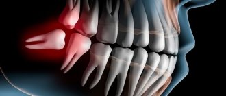
When removing the roots of wisdom teeth, you may face risks such as jaw fracture. One of the common risks of removing the roots of 3 molars is damage to the mandibular nerve
, leading to cuts and numbness in areas of the jaw. There is a risk of damaging the artery on the lingual side.
If, when a tooth is removed, a root or part of it is left behind, what should you do?
According to Russian treatment standards, the tooth root cannot be left during extraction. That is, if the patient was left with a root or part of it during removal, then this is considered a poorly performed surgical procedure.
In European countries there are doctors who agree that if there is no inflammation on the tooth and if the root part is not infected, and removal of the root leads to trauma, then only the cortical part of the root is removed, and the other part remains in the bone.
If the patient is left with a part of the root that is infected, then this is a problem. A cyst may develop and inflammation may develop. If the apical part was not infected, then everything heals and does not bring any trouble to the patient.
Tooth root without crown part
Can a tooth root without a crown cause inflammation in the gums or bone? Of course it can. There is a canal at the root of the tooth; if this canal is infected, then the problem will develop into a negative prognosis.
Inflammation, suppuration, periostitis, osteomyelitis - diseases can develop due to an infected tooth root. And the source of infection at the root must be removed; if it is impossible to remove the infection and preserve the root, then it must be removed.
Light filling on a pin – what are the advantages of the method?
A post filling is considered a reliable replacement material when a dentist is restoring a fractured unit. A titanium or fiberglass pin is inserted into the root canal, after which the coronal part is restored with a composite material. Among the disadvantages of the method is the potential risk of root fracture, since the pin fails under pressure, as well as shrinkage of the filling and the development of secondary caries under it.
Unfortunately, teeth restored in this way do not last forever. After 3–5 years, the dentist will have to change the filling or, due to global destruction of the crown, perform prosthetics/implantation.
Do these measures help stop the factors that destroy the tooth?
Any of the methods of tooth restoration involves preliminary preparation, during which the very cause of destruction is eliminated. In particular, before installing a crown, the doctor removes tissue affected by caries, cleans and seals the canals. If the specialist did it efficiently and conscientiously, as the dentists of the Unident network of clinics work, the recurrence of the disease is practically excluded. Otherwise, the patient may eventually feel pain under the denture, discomfort when chewing - all these are signs of secondary caries. In this situation, you should immediately consult a doctor - and, most likely, you will need to remove the prosthesis. And after the dentist fixes the problem that is destroying the tooth, the prosthesis will have to be made and reinstalled. That is why it is so important to turn to professionals with extensive experience and an impeccable reputation for dental restoration.
How to restore a damaged tooth?
Treatment of a damaged tooth involves the following steps:
- examination by a dentist and assessment of further “future” damaged teeth;
- restoration of the stump area using a pin or inlay. The dentist treats the canals, strengthens the unit from the inside, thereby providing reliable support for the filling;
- The crown is built up using a composite material. If the work is done efficiently, the tooth can be visually almost indistinguishable from a natural one. The dentist repeats the shape, color and even transparency.
Method of restoring teeth of the 2nd class of decay
Among the clinical cases of this category, deep carious cavities and failed fillings are most common. The main feature of the second class is that with fairly high destruction - up to 50% of the chewing surface - the main outlines of tubercles, ridges, parts of the ridges, etc. are still preserved. Having such landmarks at our disposal, we can easily continue the external contours of the preserved occlusal elements and get a chewing surface very close to the original. For this, the traditional method of layer-by-layer restoration is used, without any changes. The only addition that could help make the restoration more comfortable is the use of modeling tools in a specific sequence.
Algorithm for restoring the chewing surface. Three Tools Concept
Figure 12 shows the initial situation.
Rice. 12
Figures 13-15 show the layer-by-layer introduction of material into the cavity: a layer of dentin, a layer of chromatic enamel (the composite is distributed throughout the cavity without polymerization), a layer of surface enamel.
Rice. 13
Rice. 14
Rice. 15
To adapt the material, it is convenient to use a brush slightly moistened with adhesive (Fig. 16).
Rice. 16
Next, tool No. 1 is used - a large plugger for preliminary marking of all chewing elements (Fig. 17).
Rice. 17
They also outline the lines of future fissures and remove excess material. Modeling continues with tool No. 2 - a small plugger, which reproduces all the main forms, brings out additional ridges, side ridges, secondary recesses, and also emphasizes fissures of the first order (Fig. 18).
Rice. 18
At this stage, the excess composite is finally removed. In some cases, a small brush with lubricant can be used to make anatomical transitions smoother. The last tool (No. 3) is a sharp probe for creating the thinnest elements and emphasizing fissures (Fig. 19).
Rice. 19
After this, polymerization of the composite material is carried out (Fig. 20).
Rice. 20
As a final element, the pigmentation of the fissures can be reproduced with composite paints using a thin endodontic file (Fig. 21-23).
Rice. 21
Rice. 22
Rice. 23
Replacement of an old restoration [Clinical Case]
In (Fig. 24) the situation before treatment. An old, failing filling with obvious secondary changes in the surrounding tissue.
Rice. 24
The working field is isolated (Fig. 25) and the carious cavity is prepared (Fig. 26).
Rice. 25
Rice. 26
The quality of treatment is controlled by a caries detector. The deepest areas of the cavity were also stained, but in this case it was not the demineralized tissue that was stained, but the tertiary reaction dentin. Due to the fact that this type of dentin is formed compensatory in response to the action of irritants and is not structurally complete, has cellular inclusions and voids, it is also susceptible to indication by a caries detector. This dentinal bridge is usually the last stable barrier to the pulp chamber and should not be removed. Next, the cavity is sandblasted (Fig. 27) and adhesively prepared with the 5th generation XP Bond™ system (Fig. 28).
Rice. 27
Rice. 28
After thoroughly drying the adhesive before polymerizing it, a very thin layer of low modulus SDR™ composite was applied to the dentin and then co-polymerized. The purpose of this technique is to stabilize the hybrid zone, as well as to prevent the formation of an oxygen-inhibited layer in the adhesive interface.
Part of the cavity is filled with SDR™ composite (Fig. 29).
Rice. 29
It has a high ability to self-adapt, very low polymerization stress and optical characteristics similar to dentin. A portion of Ceram•X™ duo+ D3 composite is applied on top of the SDR™ material to imitate dentin (Fig. 30), then a layer of Esthet•X® HD YE surface enamel (Fig. 32) and a layer of Esthet•X® HD A3 chromatic enamel (Fig. 31).
Rice. thirty
Rice. 32
Rice. 31
The composite spreads over the surface, but does not polymerize. Figure 33 is a view after an instrumental simulation carried out in strict accordance with the three-instrument concept described above.
Rice. 33
Next, the characteristics were reproduced (Fig. 34, 35).
Rice. 34
Rice. 35
Figures 36, 37 show the view after occlusal integration, finishing and polishing.
Rice. 36
Rice. 37
It should, however, be remembered that in order to successfully perform a restoration using the traditional technique, the doctor certainly needs to have some experience and developed skills in anatomical modeling. The most effective way to learn to accurately reproduce the chewing surface is constant practical training.
You need to draw teeth, sculpt them from plasticine, grind them from plaster, model them from wax, reproduce them in the mouth with a composite, etc. All this must be done, guided by the general principles of shaping and knowledge of the myotopography of anatomical elements. Gradually, a sense of form will develop, the ability to “read” characteristic anatomical details, some dependencies will be revealed, and in the end a complete understanding of the entire process of reconstruction of the occlusal surface will come.
How to properly care for restored crowns?
The tooth that was restored by a specialist is no longer as reliable as a healthy one. The attitude towards him should be as gentle and careful as possible. Avoid biting hard foods; do not “test” the composite’s strength by eating dry bread or nuts. If you have restored incisors, avoid biting into hard fruits and vegetables. And remember that the fillings will have to be renewed after a few years.
Doctors at Zuub.rf perform effective restoration of damaged teeth in the smile area, as well as chewing teeth, etc. Experienced specialists will conduct an examination, prepare the crown for further restoration and guarantee the reliability and strength of the “new” tooth, which will not differ from the rest. Sign up for a consultation right now!
If you have the symptoms described in this article, be sure to make an appointment at our clinic.
Don't self-medicate! Even the smallest problem, if not treated correctly, can significantly complicate your life.
By contacting us, you can be sure that:
- Get high-quality and free consultation .
- You will receive the best prices for treatment and the opportunity to receive a special promotional price.
- Only modern equipment and materials will be used.
- You will be treated by professional doctors with many years of experience.
- We offer treatment on credit or in installments. There is also the possibility of obtaining a tax deduction.
- We work seven days a week and without a lunch break, from 9 a.m. to 10 p.m.
+7 (495) 132-02-96
Make an appointment
What is atraumatic tooth root removal?
What instruments does the surgeon use to achieve atraumaticity? For atraumatic removal, special instruments are used - thin, neat and elastic elevators that can minimally invasively penetrate and expand the periodontal tissue that connects the tooth root directly to the jaw. Using special instruments, the periodontal ligaments are cut so that the root can be removed as carefully as possible, in its “pure form.”
The vestibular plate that surrounds the bone is very thin. And we work with her carefully when removing teeth. It may also be necessary to saw the root of the tooth so that it can be removed in pieces. Sawing can be carried out with oscillating ultrasonic attachments - ultrasonic knives. Cutting the root of a tooth can be done quite effectively with a high-quality thin surgical dental bur, which has a certain length.
After cutting the root, the medial wall of the root is first removed, and then the vestibular wall of the root is removed. This allows for maximum preservation of surrounding tissue.
Then, after such removal, you can preserve the tooth socket or place an implant so that the bone is preserved as much as possible. Preservation of the tooth socket is carried out if there are no conditions for installing an implant at once, or if the patient undergoes delayed implantation.
If the roots of wisdom teeth are removed, the socket is not preserved; it is enough to remove them as atraumatically as possible. Let's talk about this in a little more detail.
Features of root removal of decayed wisdom teeth
Are there any difficulties when removing the roots of wisdom teeth? Removing the roots of wisdom teeth requires that the surgeon has sufficient experience. Often the roots of these third molars (wisdom teeth) are located close to the mandibular canal. Often the roots of molars are adjacent to the upper or lateral wall of the mandibular canal. The configuration of wisdom tooth roots is extremely diverse and sometimes extremely difficult to remove:
Therefore, when removing the roots of wisdom teeth, it is imperative that the patient undergo a CT scan. And the surgeon, as I said, needs to have some experience in surgical training in order to avoid the risks of removing the roots of such teeth and carry out the manipulation as efficiently as possible.
Like any root teeth, the roots of wisdom teeth are removed by cutting them along the roots. Wisdom teeth do not have problems because they are close to the angle of the lower jaw and there are strong cortical plates there. When I talked about thin vestibular walls, this comparison refers to the aesthetic zone of the smile, to the frontal group of teeth, including the premolars:
.
Bridges
The good old classic that dentists offer when they see that a root is missing. Bridges do not need to be removed from your mouth every day to be cleaned, unlike removable dentures, they will last 10-12 years with proper care and are much better at restoring chewing function. Modern materials are quite effective at restoring a beautiful appearance to a smile. The price and production time of such a prosthesis is higher than that of a removable prosthesis, but lower than that of an implant.
What are the disadvantages of this technique?
- With classic prosthetics relying on your own teeth, due to the uneven distribution of the load during chewing, the supporting teeth under the bridge gradually begin to collapse.
- Bone atrophy also does not stop.
- The denture requires special cleaning products - pipe cleaners, brushes, etc., otherwise food debris and plaque will accumulate under it.
- The implant-based method is not always possible - if one tooth or two in a row is lost, the doctor will not remove adjacent healthy teeth in order to put implants in their place.
What to choose?
What to install if there is no root - a removable denture, a bridge or an implant? The dentist will select prosthetic options based on the condition of the oral cavity and the presence of relative or absolute contraindications. However, today doctors most often offer implant prosthetics. It has only two disadvantages compared to other types - a higher price and a longer period of prosthetics with several stages. However, this is compensated by excellent, long-term and predictable results. In addition, implantation technologies are constantly being improved; there are already techniques and implants that can significantly reduce the time of prosthetics.
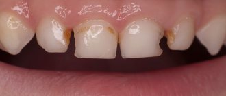
Crack on front tooth
The front teeth always get the most damage. When they “punch you in the teeth,” the blow usually falls on them. They fell unsuccessfully, worried, chewed a pencil, or, horror of horrors, chopped nuts with their incisors, and now a crack appeared on the front tooth. By the way, it can occur without mechanical damage - for example, due to temperature changes. Teeth don't really like iced coffee. You shouldn’t overuse citrus fruits or their juices either, and it’s best to dilute freshly squeezed juices with water to reduce their acidity. You should not get carried away with whitening procedures, and be more careful with abrasive pastes. Cracks are also different.
Vertical
If you are “lucky” and it is small vertical, then nothing bad will happen to this tooth, and the defect will be barely noticeable. In this case, many dentists do not recommend loading the tooth with patches. But you still need to keep an eye on the crack. If it increases, the enamel changes color, and the tooth reacts sharply to hot/cold, sweet/salty, you should consult a dentist. The enamel will need to be restored and strengthened. The crack will be treated with a special compound containing calcium and fluorine. After the course of treatment, the tooth will react normally to food and become more even and smooth.
Horizontal
But horizontal or oblique cracks in the front teeth are a cause for serious concern. They provoke the formation of caries, chips and can generally lead to tooth loss. Treatment depends on the size, depth and location of the crack, as well as the characteristics of the enamel. If there are many microcracks on the tooth and the color of the enamel has changed, the doctor may suggest installing veneers, but only after treating caries and other oral diseases. A crack left unattended will permanently split the tooth. Then there will only be one treatment option left - prosthetics.
Prevention of tooth decay in adults
Careful care will help keep your teeth healthy. It consists of a number of factors, including concerns about nutrition and lifestyle.
In addition, it is important to purchase:
- toothbrush, soft or medium hard. Do not forget that the brush should be changed regularly;
- threads and rinsing gels;
- toothpaste selected after consultation with a dentist.
Bad habits are worth fighting. This applies not only to smoking or alcohol, but also to clicking teeth, chewing crackers, pistachios and other hard foods.
If involuntary teeth grinding occurs, you will need the help of a neurologist. In most cases, the solution to the problem is mouthguards and sedatives (they are used only as prescribed by a doctor).
Try to enrich your diet with seafood, fresh vegetables, cottage cheese, and fruits. For rinsing (and it is better to do it after each meal), use special products. And don’t forget about the importance of preventive examination: you need to visit the dentist once every six months.
Number of views 6,832
Is implantation possible after tooth root removal?
Yes, of course it is possible. And we at the clinics of the German Implantology Center widely use implantation after tooth root removal. Our conditions allow us to carry out implantation in such cases. And if we can achieve primary stability of the implant, then in 90% of cases
After removal, we immediately install the implant. We have rolled out the technology to perfection and gives excellent results.
Of course, with the exception of wisdom teeth, no implant is placed after their removal. But, by the way, wisdom teeth in this situation can be extremely useful, since they... can be transplanted to the place of the problem tooth being removed! Dental transplantation has been practiced in the clinics of the German Implantology Center since 2017, and our specialists have accumulated a wealth of clinical experience. Let's talk about this a little more.
Content
- Dental restoration methods
- Dental restoration with pins
- Dental restoration with inlays
- Dental restoration with crowns
The success of endodontic treatment largely depends on the subsequent restoration of the upper part of the tooth. It is the correct restoration of the crown after filling that is the key to a long-term, high-quality result without the risk of re-infection. Canal treatment is not considered complete until the doctor can guarantee that there is no risk of re-contamination.
Treatment methods – from filling to implant
The dentist will select the treatment method for the front teeth after an examination.
- Veneers or Lumineers
If the incisors are severely damaged, thin overlays can be placed - veneers or lumineers. They will hide the consequences of caries and make teeth whiter and smoother.
- Dental crowns
If only the root remains of the front tooth, intra-root inlays are made; this is the basis for the crown. Metal crowns are not suitable for front teeth; they will be too noticeable. To make an artificial tooth as similar as possible to a real one, it is better to use ceramic and zirconium crowns.
- Bridges and implants
If desired, missing front teeth can be replaced by bridges and clasp dentures. If funds allow, you can install an implant and forget about the problem for many years.
Why do teeth rot?
- Poor or no oral hygiene
- Hereditary factor
- Incorrect diet (too much flour, sour and sweet foods)
- Bad habits (alcohol, cigarettes, drugs)
- Caries
- Inflammatory processes in the oral cavity
- Lack of beneficial vitamins and minerals in the body
- Hormonal imbalance (quite often teeth rot during pregnancy and lactation, as well as during adolescence)
- Stomach problems
- Thyroid diseases
- Bad ecology