Abrasion is the process of loss of hard dental tissues. Tooth wear occurs both in the temporary and permanent dentition; both occlusal and proximal surfaces; both at reduced speed and at increased speed. Depending on the severity of this process, physiological and pathological abrasion are primarily distinguished.
As a result, pathological abrasion of teeth occurs
The pathogenesis of the disease involves several factors, both external and internal.
| Endogenous factors | Exogenous factors |
| Metabolic disease. Gastrointestinal diseases. Pathology of the endocrine system. Bruxism (teeth grinding). | Patient's nutrition. Nature of working conditions. Incorrect prosthetics. |
The greatest influence on the abrasion process itself is exerted by occlusal forces, which have the following characteristics:
- The amount of chewing force. It is determined by the strength of the masticatory muscles, which can range from 80 to 400 kg. It is necessary to take into account that teeth are under stress not only when chewing food, but also when swallowing, and a person performs several thousand swallowing movements per day.
- Duration. During the day, the occlusal surfaces of the teeth come into contact with each other for half an hour, but with pathology, this figure can increase significantly.
- Application point and direction. The occlusal force is directed at right angles to the chewing surface of the tooth. Normally, the contact area is several square millimeters, but with pathology it can increase several tens of times, which leads to greater efforts when chewing.
In the presence of harmful production factors, bruxism, fluorosis and other reasons mentioned above, all characteristics of occlusal forces change for the worse, which leads to tooth abrasion.
Non-carious lesions of hard dental tissues: to treat or not to treat?
The loss of dental hard tissues or their abrasion is a process that can be characterized by both physiological and pathological development patterns, which ultimately leads to the formation of non-carious dental lesions. The latter can compromise the aesthetic profile of the smile, disrupt the function of the dentofacial apparatus, provoke sensations of pain and sensitivity, and negatively affect the patient’s quality of life. In the United States, the problem of non-carious dental lesions has not been studied as well as in Europe or Australia, so a unified protocol for their treatment has not yet been developed, which would be based on the available evidence. Overall, the more important question has not even been answered: should such lesions be treated or not?
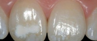
The structure of the teeth is represented by mineralized tissue, which dynamically undergoes the process of mineralization and demineralization when exposed to oral factors. In the normal state of saliva, calcium and phosphorus ions are washed out to a greater extent from the tooth structure, but when saliva is saturated with fluorides, the reverse process occurs, which ensures homeostasis. Under the influence of chemical and mechanical factors, there is a loss of hard tooth tissue, which can be presented in the form of erosion, abrasion, attrition, abfraction and permanent loss of mineral components by enamel and dentin (photo 1).
Photo 1. Type of physiological tooth wear.
Loss of surface tooth structures due to microbiological activity leads to the development of carious lesions. In turn, the dissolution of mineral components under the influence of chemical agents or chelates from the tooth surface not covered by dental plaque is called erosion. Erosion is not directly related to the action of mechanical or traumatic factors, as well as to carious lesions. However, erosions are often identified together with signs of attrition, abfraction and abrasion. Attrition is the loss of tooth structure due to physical contact between opposing teeth, abrasion is the result of physical abrasion involving abrasive materials, and abfraction is due to tensile and shear stresses, which are usually noted in the cemento-enamel region. If there are signs of several non-carious pathologies at once, it is diagnostically difficult to establish the main cause of the loss of hard tooth tissue.
Classification of non-carious lesions of hard dental tissues
There are four main types of non-carious lesions of hard dental tissues, but in most clinical cases there are signs of several of them at once, which complicates the process of making a final diagnosis.
Erosion
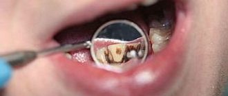
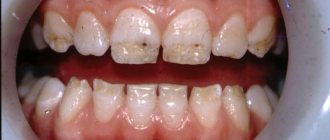
Erosive lesions of the hard tissues of teeth occur due to the effects of acidic or specific erosive factors of the internal or external cut, the intensity of which exceeds the buffering capabilities and neutralizing potential of normal saliva and its proteins. The term “corrosion” also exists to refer to the loss of enamel and dentin tissue as a result of chemical and electrochemical factors. Dental erosion develops when the level of demineralization of calcified hard tissues exceeds the level of remineralization of them. The dissolution of enamel hydroxyapatite crystals occurs at pH 5.2, and of dentin at pH 6.9. Under normal conditions, the tooth structure remineralizes with the formation of hydroxyfluorapatite crystals, which are more resistant to acid. But be that as it may, the duration and frequency of acid exposure determine the risk of developing erosive lesions. Saliva is the most significant biological factor that can prevent the development of erosion. Neurological communication mechanisms stimulate saliva production when oral acidity levels increase, thereby increasing the buffering potential of saliva, dissolving concentrated acids, and maintaining oral homeostasis. The supersaturated concentration of calcium and phosphate in saliva promotes the remineralization of demineralized tooth surfaces. Saliva also plays a role in the formation of the pellicle, which acts as a barrier to protect tooth surfaces. Factors that help prevent erosion also include the tooth structure itself and the position of the tongue. Signs of carious lesions of hard tissues are a decrease in the height of enamel and dentin from the occlusal surface of molars, and the formation of saucer-shaped defects on the tubercles of the latter (perimolysis). With erosion, both minimal signs of damage can be observed, such as loss of shine of the teeth, a dull or matte appearance of the surface, as well as severe symptoms associated with the loss of hard tissues to the level of dentin. As a rule, the width of eroded areas is greater than their depth. When erosive lesions are combined with abrasive ones, the loss of enamel and dentin occurs more quickly and progressively (photos 2-5).
Figure 2: Loss of hard tissue around amalgam restorations.
Photo 3. Appearance of non-carious lesions in patients with GERD.
Photo 4. Combination of erosion and abrasion.
Figure 5. Hard tissue loss and yellow discoloration of teeth in a young patient with GERD.
Abrasion
Abrasion is the loss of hard tooth tissue due to friction (exposure) to abrasive agents. Such lesions occur when toothpaste and brushes are used incorrectly. The severity of abrasion depends on the level of abrasiveness of the active agent, the magnitude of the acting force, the time of exposure, and the frequency of development of physical contact (photo 6). Abrasives in the structure of toothpastes include silicates, aluminum, calcium phosphates and calcium carbonates. Typically, the abrasive changes caused by brushing are minimal, but this depends on the type of brush used and the concentration of abrasive in the toothpaste. The abrasiveness of the toothpaste suspension affects the level of hard tissue loss, therefore, the composition of the toothpaste must be taken into account when choosing it before use. However, some studies found no correlation between the composition of toothpaste and the development of cervical lesions of the teeth. Abrasive lesions are characterized by a larger axial depth parameter than the occlusal-gingival width, and sharp angles are often noted in their geometric structure.
Photo 6. Erosion and abfraction in a patient with xerostomia in a photo of Sjögren's syndrome.
Attricia
Attrition is the loss of dental hard tissue due to mechanical contact with opposing teeth, or with restorations on opposing teeth. When occlusal relationships are violated, signs of attrition become visible on natural teeth that interact occlusally with restorations, since the latter are harder than the structure of intact enamel and dentin. Often, signs of attrition are most pronounced in the area of occlusal and contact surfaces, as well as in the projection of the cutting edge of the teeth. The severity of attrition is influenced by the type of chewing, swallowing and the presence of parafunctional activity. In the absence of control of these factors, abrasion facets develop in the enamel projection, which have a flat and clearly defined shape on both antagonist teeth (photo 7).
Photo 7. Metal-ceramic structures that antagonize the patient’s own teeth.
Abfraction
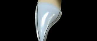
Abfraction is the result of static and cyclic loading, which develops due to the action of various biomechanical forces on the tooth structure. The effects of biomechanical forces that act on the weakest part of the tooth, that is, the cemento-enamel junction (CEJ), depend on the magnitude, duration, direction, frequency and location of the forces. As a result, the enamel or dentin can chip, forming non-carious lesions in the neck of the tooth. The latter are characterized by a specific V-shape (photo 8). Most data on abfraction are based on “engineering” models of studying the tooth, or separately its coronal and root parts. However, more in-depth studies of the function and interaction of the periodontal ligament and alveolar bone have called into question the fundamental theoretical basis of the theory of abfraction development associated with the influence of biomechanical components.
Photo 8. V-shaped cervical defects of teeth as a result of occlusal overload.
Common causes of the development of non-carious lesions of hard dental tissues
Bruxism / clenching
Bruxism is a pathological habit of non-functional clenching and further movement of the jaw (parafunction) into an eccentric position, which includes the canine and incisal routes of insertion. Bruxism is associated with significant hard tissue loss (three to four times the normal rate of wear ranging from 10 to 20 µm per year), which leads to structural changes in tooth morphology with the formation of deep grooves and wear facets. Clanching is considered a “silent form of bruxism” in central occlusion without lateral or protrusive movements of the jaw. The loss of hard tissue in the area of the anterior teeth is much more pronounced than in the area of the distal teeth.
Pathology
Diseases that provoke the loss of hard tooth tissues include pathologies associated with disruption of the natural process of formation of enamel and dentin (impaired emalogenesis, impaired dentinogenesis). With a decrease in the mineral density of the tooth, the level of mineralization and hardness of the structure, the teeth become less resistant to the effects of physical and chemical factors. In addition, the level of oral defense reactions decreases when the normal function of saliva is disrupted due to its deficiency or dysfunction of the salivary glands. These conditions are associated with such general somatic pathologies as Schoengren's syndrome, rheumatoid arthritis, as well as with radiation therapy in the head and neck area. With hypofunction of the salivary glands, there is a deficiency of saliva, which in turn provokes an increase in the level of friction between the teeth, as well as between the teeth and the toothbrush. Locally specific loss of hard dental tissues is a protective mechanism, or a compensatory effect of the structures of the dentofacial apparatus. Gastroesophageal reflux disease (GERD) also has a negative effect on the structures of hard tissues, due to which the pH of the oral cavity is significantly reduced, which leads to the dissolution of mineral components in the tooth structure. Tooth erosion can be considered an atypical symptom of GERD. Voluntary regurgitation is also observed in cases of anorexia nervosa and bulimia, which similarly leads to acid damage to the teeth. Various medications with anticholinergic side effects (eg, psychiatric medications, antihypertensives, drug combinations, etc.) cause decreased salivary flow. Chronic use of drugs with low pH (solutions, chewable tablets or inhalations) has a detrimental effect on teeth and can potentially provoke the development of erosive lesions of enamel and dentin.
Diet and lifestyle
Frequent consumption of acids, especially citric and acetic acids in various drinks and foods, provokes the dissolution of minerals in the structure of teeth and reduces their microhardness. pH is not the only parameter influencing the resistance of hard dental tissues. The integrity of enamel and dentin structure is also dependent on the erosive potential of foods and beverages, the buffering properties of saliva, and the calcium chelation pattern recorded with different types of diet. Foods and drinks containing citric acid are characterized by a more pronounced erosive effect than those containing phosphoric acid. Diet drinks that are low in calories and sugar do not actually provide adequate caries prevention because their erosive potential causes the hard structures of enamel and dentin to dissolve and thus increase their susceptibility to bacterial attack. A high risk of damage to hard tissues is characterized by the consumption of “sports” drinks with a high level of acidity (pH = 3), especially immediately after sports in conditions of dehydration of the oral cavity. On the other hand, consuming hard foods that require careful chewing also increases the risk of developing pathological loss of hard dental tissue. Personal habits such as smoking a pipe, chewing a pen or pencil, or opening bottles with teeth also provoke the development of non-carious lesions. Working under conditions of constant stress, in factories with a high level of air acidity, in noisy conditions, swimming in chlorinated pools, professional wine tasting, and playing certain musical instruments are all factors in the medical history that can negatively affect the condition of the structures of the hard tissues of teeth. .
Brushing teeth and toothpastes
Brushing teeth that are in a demineralized state after exposure to acid causes a cumulative effect of loss of hard enamel and dentin tissue. Brushing teeth in a horizontal direction causes more wear on the cervical portion of the tooth compared to a vertical brushing pattern. In addition, the type of bristles of the toothbrush, as well as the type of toothpaste used, have a pronounced mechanical effect on the surface of the teeth. More pointed filaments of a toothbrush, a larger number of tufts, their dense arrangement and stretching under load are the factors that most likely negatively affect the abrasion of hard dental tissues and restorative materials. A certain level of abrasiveness in toothpaste is necessary to effectively remove bacterial biofilm and stains from accessible tooth surfaces, as well as to polish them during the brushing process. However, the levels of abrasiveness of toothpaste, determined by the RDA and REA methods, are significantly associated with the risk of abrasion of tooth surfaces, therefore, these indicators must be paid attention to when choosing a toothpaste. Interestingly, at the legislative level, RDA indicators may not be indicated on the product packaging; in this case, it is better to check the exact parameters on the manufacturer’s website.
Craniofacial complex
Vertical deviations, a small angle between the mandibular and palatal planes, as well as a small angle of the lower jaw can affect the level of abrasion of tooth surfaces. The forces and tensions that develop during chewing can reach quite high values, since the masticatory muscle, responsible for their development, is one of the strongest in the human body. The more square the patient's face looks, the larger the area of attachment of the masticatory muscles, which means the greater the bite force and pressure. It is logical that with stronger biting, the risk of pathological loss of hard tissue increases markedly.
Iatrogenic dental causes
In cases where the hardness of restorations or crowns exceeds the hardness of their own antagonist teeth, signs of pathological abrasion begin to develop in the area of the latter. To avoid these, it is necessary to carefully monitor the levels of occlusal relationships. Typically, problems with pathological abrasion of teeth are localized and projected onto teeth that antagonize different types of orthopedic structures, although in some cases they can also be generalized.
Aging
Abrasion of hard dental tissues occurs physiologically and with age. During the functioning of the teeth, the tubercles are smoothed out, wear facets are formed, the volume of enamel decreases and the dentin surface is exposed. As the life expectancy of the population increases, the period of dental function also increases, which is why the prevalence of gum recession is increasing, exposing areas that are normally located below the level of the gums. Among older patients, whose teeth have been functioning for a longer period of time, the risk of developing erosive non-carious lesions increases against the background of the development of recessions.
Consequences of non-carious lesions
The consequences of non-carious lesions depend on the degree of clinical severity of such lesions. Firstly, it is possible to lose the entire enamel structure with exposure of the dentin surface, changes in the transparency of the tooth and yellowing of its structure. Secondly, wear facets may form on the occlusal surfaces and flattening of the cusps may be noted. When the dentin surface is exposed, patients often complain of hypersensitivity, but in some patients, on the contrary, sclerosis of dentin and its tertiary deposition develop, therefore, there are no signs of hypersensitivity. A decrease in the height of clinical crowns due to pathological abrasion is also fraught with a reduction in the height of the bite, which negatively affects the function of the temporomandibular joint. Progressive pathological tooth wear is also associated with the risk of developing pulpal lesions, and in the frontal areas - with a compromise in the external profile of the smile. It is logical that in such conditions, patients want to restore the natural appearance of their teeth and solve existing functional problems on the part of the dentofacial apparatus, and all this entails a set of financial costs. Thus, non-carious dental lesions are associated with a violation of the parameters of function and aesthetics, and, accordingly, with a decrease in the overall quality of life of patients.
Treatment
Treatment of non-carious lesions of teeth should be comprehensive and take into account the action of the main etiological factors. A simply restorative approach to restoring lost tissue without taking into account the root causes of the damage is often unsuccessful. So far, no evidence-based recommendations have been developed regarding the prevention of the development of non-carious pathologies of hard dental tissues. But the dentist must be well aware of the reasons for their development in order to inform the patient about all treatment options, its prognosis and features.
Preventive methods
To assess the effectiveness of preventive programs aimed at preventing the development of non-carious lesions of enamel and dentin, it is necessary to record the condition of teeth using special cards, photo registration, surface mapping and model analysis. It is primarily recommended to gradate the level of severity of pathological abrasion. According to the Tooth Wear Index 0, the level of hard tissue loss corresponds to a stable state of enamel characteristics (B/L/O/I) and an integral tooth contour (C); Level 1 – loss of enamel characteristics (B/L/O) and minimal loss of tooth contour (C); Level 2 – loss of enamel and dentin exposure on one third of the tooth surface (B/L/O), or loss of enamel with minimal dentin exposure (I), or a contour defect up to 1 mm deep (C); Level 3 - loss of enamel and dentin exposure of more than one third of the tooth surface (B/L/O), or loss of enamel with significant dentin exposure (I), or a contour defect 1-2 mm deep (C); Level 4 – complete loss of enamel, exposure of pulp or secondary dentin (B/L/O), or exposure of pulp and secondary dentin (I), or contour defect more than 2 mm deep (C). According to another classification, class I of non-carious pathologies includes vestibular lesions in the early stages of erosion with the absence of obvious contour changes, a smooth shiny surface mainly on the labial side of the upper incisors and canines. Class II lesions involve the involvement of dentin at 1/3 of its depth (type 1: ovoid or cup-shaped lesions of the cervical areas, which must be differentiated from wedge-shaped defects; type 2: uneven lesions of the entire crown). Class IIIa lesions include those with more extensive destruction of dentin, especially in the area of the anterior teeth, and the lesions themselves are characterized by a fairly large area; at the same time, class IIIb lesions extend to the lingual surfaces and cutting edge, and the lesions themselves are characterized by the presence of smooth dentin. In class IIIc, the cutting edges and occlusal surfaces are affected down to the dentin levels and become flattened; existing restorations may even protrude somewhat above the level of their own tissues; the cutting edges appear overly transparent. Class IIId lesions characterize pronounced disturbances in the structure of the teeth on both the vestibular and lingual sides.
By monitoring changes in the lesion according to the classifications described above, it will be possible to monitor how effective a particular method of prevention and treatment of non-carious pathologies is. In addition, it is necessary to pay sufficient attention to general somatic pathologies that may be associated with loss of enamel and dentin of non-carious etiology (GERD, diseases of the salivary glands, chronic renal failure). If there is a deficiency of saliva, you can use special stimulating drugs (pilocarpine HCl or cevimeline HCl). In cases of xerostomia developing while taking certain medications, they should be replaced with others with a less pronounced indirect cariogenic effect. Diet is also an important component of the prevention program. Patients should keep a food diary for a week and review it to minimize the intake of acidic foods and switch to less acidic foods fortified with fluoride and minerals such as calcium and phosphate. It is also recommended to eat neutralizing foods, such as cheese. To normalize pH levels after meals, it is advisable to rinse your mouth with plain water, a low-fluoride mouthwash, milk, or baking soda (sodium bicarbonate). At the same time, you should not start brushing your teeth immediately after first eating acid-containing foods; first, it is better to rinse your mouth in order to normalize the pH. It is also recommended to use less toothpaste and toothpastes with low relative abrasiveness in general. Patients should also be told about the possibilities of modifying their brushing technique to use less force and the benefits of using brushes with rounded bristles. In the presence of bruxism, the treatment protocol becomes more complicated, and it should, among other things, include changing some behavioral aspects and habits, such as, for example, correcting the influence of stress factors, studying relaxation therapy methods (yoga, self-relaxation, self-hypnosis). In certain cases, the doctor may prescribe the use of a mouthguard to mechanically separate the tooth at night or during the day.
Remineralization of tooth structure
Every effort should be made to ensure effective remineralization of the tooth structure and to minimize the progression of hard tissue loss of enamel and dentin. Fluoride promotes the reuptake of available calcium and phosphate present in saliva. However, fluorides are available in various concentrations and can be used by the patient, for example, in the form of sodium fluoride (in the form of toothpaste with a concentration of 5000 ppm), or applied to the teeth by the dentist during a visit (in the form of varnish with a concentration of 26,000 ppm). Mineral reuptake can be improved by adding or using calcium and phosphate ions in various forms along with fluoride. In turn, fluorides, sodium hexametaphosphates, ferrous sulfate and fluorinated tin rinses also act as anti-erosion agents.
Dental restoration
Physiological tooth wear does not always require treatment. However, if the process of hard tissue loss is pathological, then the patient should be given appropriate treatment. Active treatment depends on the individual characteristics and needs of the patient, the extent of the lesion, the skill of the dentist, time available and cost. Evidence based on dental analysis in accordance with generally accepted engineering principles indicates that non-carious lesions should be restored. To do this, it is first necessary to ensure control over the etiological factors to ensure the success of all subsequent manipulations. Restoration of cervical non-carious defects requires special attention, taking into account the development of all forms of stress that develop in the tooth during occlusal interaction. When restoring cervical defects, the amount of preparation should be kept to a minimum to ensure only the roughness of the affected surfaces. The most common cause of unsuccessful restoration of cervical non-carious defects is microleakage. In this case, it is advisable to carry out restorations only when the positions of central occlusion and the central ratio have converged into one spatial position, or when a state of compensation has been achieved in the structure of the dentofacial apparatus. Restorations for non-carious lesions include those with overlapping tooth surfaces with composite material using the sandwich technique, as well as designs of metal, metal-ceramic and ceramic crowns (photo 9-16).
Photo 9. Non-carious lesions of teeth according to class 5.
Photo 10. Type of non-carious lesions of class 5 restored using composite restorations.
Photo 11. Compromise of the aesthetic appearance of teeth as a result of pathological abrasion.
Photo 12. View of the patient’s teeth after restorations have been completed.
Photo 13. Compromised aesthetic appearance of teeth due to pathological abrasion.
Photo 14. View of the patient after rehabilitation with metal-ceramic structures.
Photo 15. Type of non-carious dental lesions in a patient with bulimia.
Photo 16. Restoration of defective areas with composite restorations.
If the central occlusion does not correspond to the centric relation, and there are signs of localized pathological abrasion in the area of the lower anterior teeth, it is first necessary to ensure careful monitoring of the condition of the dentofacial apparatus. If signs of localized pathological abrasion are visualized in the area of the upper teeth, restorations can be performed and a Dahl device can be used. The latter is a metal bite plate, which is used to restore areas of teeth affected by pathological abrasion by forming the necessary interocclusal space. When the intermaxillary space increases due to loss of bite height, it is first necessary to establish the position of the central relationship, and only then proceed to dental restorations. When the vertical height of the bite increases, it is recommended to use stabilizing mouth guards. If the patient can normally tolerate the height of the aligner, then in such cases restorations can begin; If the patient cannot tolerate the height of the aligner, then the clinician should consider using an approach of lengthening the height of the clinical crown.
conclusions
Pathological abrasion of teeth and associated non-carious lesions of enamel and dentin are pathologies that are difficult to objectively diagnose with verification of the main cause of the disorders. The etiology of noncarious lesions is individual in each individual clinical situation, and the signs of this disorder vary between patients in terms of both patterns and types of hard tissue loss (erosion, attrition, abrasion, abfraction, or a combination of these). To ensure a successful outcome in the treatment of pathological abrasion, the doctor must take into account the etiology of such, the possibility of implementing preventive approaches and targeted therapeutic interventions, based on the conditions of each individual clinical case.
Authors: Mabi L. Singh, DMD, MS Gerard Kugel, DMD, MS, PhD Athena Papas DMD, PhD Britta Magnuson
When to start treatment
The problem does not appear suddenly; it is characterized by a long course with gradual progression. The sooner treatment begins, the shorter and less radical it will be. That is why you should contact your dentist when the first signs of the disease appear, which include:
- Increased sensitivity of dentin.
- Reaction to hot and cold food.
- Changes in the color and shape of teeth.
- Early abrasion of enamel.
It must be remembered that there is also physiological abrasion of teeth. It is characterized by a long and slowly progressive course and is a consequence of the natural aging processes of the body. If a patient experiences rapid wear of enamel and dentin at a young age, then it is necessary to immediately contact a specialist for professional help.
How is pathology determined?
Only a specialist can detect a violation during tooth wear. Of course, the general picture among the population is taken as the norm, but the doctor also takes into account individual factors:
- general condition of the enamel;
- presence or absence of exposed dentin;
- work of the temporomandibular joint;
- condition of the mucous membranes of the tongue and cheeks;
- degree of severity of nasolabial folds;
- sensitivity of the masticatory muscles.
It is interesting that in addition to all the individual characteristics, the doctor is interested in the sound with which the jaws close. A sharp, clear and short sound indicates that there are no problems. A long-lasting sound accompanied by a squeak indicates deficiencies in the functioning of the TMJ or problems with the nervous system.
You can understand that teeth are subject to increased abrasion by the first sign - increased sensitivity. It is sharper when the upper hard layer of enamel is significantly abraded.
Patient examination plan
Traditionally, the examination begins with getting to know the patient, collecting anamnesis and complaints. At this stage, the doctor finds out when the disease began, how it manifests itself, what it may be associated with (the patient’s lifestyle, previous treatment, harmful occupational factors, etc.). If the problem has just begun to manifest itself, then complaints as such may be absent or only slightly expressed. Typically, patients note discomfort when eating hot and cold food, chipping of the enamel or its thinning, and changes in diction (in later stages of the disease).
At the next stage, the doctor proceeds to examine the oral cavity, during which he evaluates:
- Bite.
- Overlapping of incisors.
- The shape of the dentition.
- The shape of the teeth.
Based on the data obtained, the doctor can prescribe additional research methods (radiography, MRI, electroodontodiagnostics, etc.), as well as consultations with more specialized specialists.
Treatment methods
Treatment of pathological tooth wear may include therapeutic and orthopedic techniques. In the first case, there are two options:
- Drug therapy (includes the application of gels and solutions, the use of special toothpaste).
- Restoration (special filling material is used).
Orthopedic treatment involves the manufacture and installation of onlays, dentures or crowns, which restore the normal appearance and functionality of teeth.
Currently, dentists do not agree on the timing of initiation and specific treatment methods in different situations. Most often, drug treatment is used in patients in the early stages of the disease, when the teeth are not yet seriously damaged. For more severe pathology, restorative and orthopedic treatment is indicated.
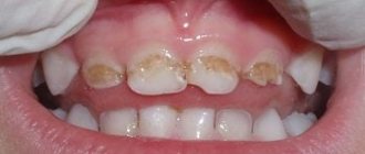
What to do if your child's teeth are worn out
Treatment for tooth wear is developed individually. At the appointment, the pediatric dentist analyzes the causes of the pathology, determines the stage of erasure, the nature of the disease and the characteristics of the development of the child’s body.
Restoration of worn teeth
Restorative treatment is carried out while preserving at least half of the hard tissues of the tooth. The technique takes place in several stages:
- Preparation. Necessary to identify the causes of diseases and their elimination or minimization, as well as the protection of remaining teeth. During preparation, an occlusal splint is made and installed if the patient suffers from bruxism. If necessary, the doctor prescribes consultations with various specialists, explains the rules of oral hygiene, and sanitizes foci of infection.
- Evaluation of the preparatory stage. After some time, the patient comes for an appointment, and the doctor assesses the condition of the teeth, checks whether the patient followed all the recommendations correctly, and discusses the upcoming treatment with him.
- Restoration. It is carried out in a certain order. First, the teeth are prepared, then the teeth on the upper jaw are restored, and only then on the lower jaw. Using a table of anthropometric data for each tooth, the doctor gives the patient's new teeth a certain length and shape.
- Observation. After successful treatment, the doctor observes the result for some time. In the first year, the patient should come for an appointment once a month, then less and less, but not less than once every 12 months. If any defects are identified, they are repaired or other treatment methods are applied.
This type of treatment, like any other, requires the patient to strictly adhere to all recommendations, give up bad habits, eat right, regular examinations by a specialist, and carefully observe hygiene skills.
Treatment of tooth canals before prosthetics
The healing of the implants was successful, so immediately after removing the braces, it was possible to begin dental prosthetics. Before dental prosthetics with crowns, the dental canals were re-treated with a microscope and the teeth were strengthened with pin inlays, where required. It is absolutely not necessary to depulpate teeth (treat canals) in all teeth on which crowns will be installed. Experienced Dial-Dent dentists install crowns on living teeth, using special tooth preparation technologies.
Crown as a way out
Pathological tooth wear can be eliminated with the help of crowns. They can be installed both on the front teeth and on the chewing teeth and restore the aesthetics and functionality of the oral cavity. Crowns can be made from various materials, each of which has its own advantages and disadvantages:
| Material | pros | Minuses |
| Metal ceramics | Durability, high aesthetic qualities, not the highest price | Depulpation of the tooth and grinding of a large amount of hard tissue are required. |
| Metal-free ceramics | The best aesthetics that are not lost over time, high strength, durability. | High price. |
| Metal | High strength, durability, low cost. | Lack of aesthetic qualities. |
When choosing a certain material, the patient must understand that he will need to replace a large number of crowns (perhaps even all). Therefore, it is worth giving preference to those materials that have good aesthetic qualities and a long service life.
After all the nuances of prosthetics have been agreed upon, we proceed directly to the procedure itself. At the first stage, the oral cavity is sanitized and teeth are prepared for crowns. Next, the doctor takes impressions of the jaws, based on which the dental technician makes a prosthesis. These operations may take some time, so the patient may be offered temporary plastic crowns. They will protect prepared teeth from the aggressive environment of the oral cavity and help adapt to new teeth. After the final version of the prosthesis is ready, it is tried on again and fixed with permanent cement.
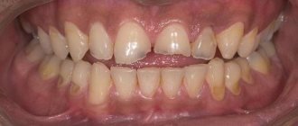
Consequences and symptoms
Pathological abrasion threatens to expose the tooth pulp and increases the risk of the appearance and development of a carious cavity. This can lead to pulpitis. The pulp, in turn, activates its protective mechanisms, which manifest themselves either in the formation of secondary dentin.
Pathological abrasion leads to tooth deformation, which in some cases requires its removal. To save the affected teeth, treatment must begin immediately. In addition, the pulp may die as a result of the fact that replacement dentin simply does not have time to be deposited
The manifestation of increased abrasion traditionally begins: a painful reaction of the teeth to cold and hot. The teeth react with the same sharp pain to chemical irritants. If the secondary layer of dentin still managed to protect the pulp, then increased sensitivity of the teeth may not be observed.
If the pulp has already died, foci of inflammation can be found at the tops of the roots of the teeth.
Restoration and normalization of the dental system
These tasks are implemented during orthodontic treatment. The correct position of the teeth and physiological bite not only improve the aesthetic function of the oral cavity, but also contribute to the uniform distribution of the load on the teeth. To achieve such results, orthodontic appliances are used, which can be removable or non-removable. In the first case, the Dahl method is used, the essence of which is that a pre-made bite pad is installed on the inner surface of the front teeth, which is necessary for uniform distribution of pressure vertically. The platform is fixed with clasps to the supporting teeth (usually canines and premolars); if necessary, the patient can remove it independently.
Fixed appliances are permanently fixed in the oral cavity for a long period of time; they are secured with cement, so the patient cannot remove them. The most popular are the Angle apparatus, the Ainsworth apparatus, the Simon apparatus, etc. Correctly selected orthodontic treatment can make the upcoming restoration or prosthetics simpler and more effective.
Modern materials and treatment methods in dentistry make it possible to successfully combat pathological tooth wear. The interaction between doctor and patient is very important. The specialist must choose the right treatment method and give the patient all the necessary recommendations, which he must strictly follow. The responsibility of both parties will be a good guarantee of a successful fight against the disease.
Dial-Dent specialists who took part in the treatment:
- Orthopedic dentist S.V. Zukor – drawing up a comprehensive treatment plan, coordination of treatment, dental prosthetics with ceramic crowns.
- Orthodontist M.P. Sleptsova – treatment with braces: elimination of crowding of teeth, correction of bite, preparation for prosthetics on implants.
- Implant surgeon V.P. Alaverdov – installation of Astra Tech dental implants, sinus lifting, bone grafting, removal of wisdom teeth.
- Dentist-endodontist T.I. Matienko – treatment of tooth canals with a microscope, replacement of old fillings, treatment of caries.
- Dental neurologist E.V. Saxonova – consultation on the issue of night grinding of teeth and severe clenching of teeth (bruxism).
- Dental hygienist T. Kondratieva – hygienic preparation for treatment, hygienic support during treatment.
- Dental technician D.V. Wolf – production of ceramic dental crowns and veneers.
- Dental assistants M. Erkimbekova, S. Shchelkova, L. Kharlamova, A. Antoshkina.
The cost of treatment depends on the specific situation, so treatment at Dial-Dent begins with an examination, diagnosis and drawing up an estimate for treatment, where all the necessary procedures and their prices are prescribed. The administrator will not be able to tell you the price of treatment over the phone, since the price in the price list is only the price of individual treatment procedures. Without examination and diagnostics, it is impossible to say what exactly and in what volume will be required. Consultation with a dentist at Dial-Dent is free, you may only need to pay for the images (sight image - 390 rubles, panoramic image - 1300 rubles, computed tomography of the teeth - 2900 rubles). Only after signing the estimate, when the patient understands what will be done and how much it costs, treatment begins.
See other examples of treatment from Dial-Dent specialists here.
Make an appointment for a consultation by phone +7-499-110-18-04 or through the form on the website. You can ask questions about dental prosthetics to the chief doctor of the clinic, Sergei Vladimirovich Tsukor, at