When a child complains of acute pain in the ear, cries and is capricious, this state of the baby will unbalance even the most persistent and experienced parent. Acute shooting pain in the ear is not easy for an adult to endure, let alone for children.
Pain signals the appearance of an inflammatory process in the ear. That is, otitis media develops. According to statistics, by the age of five, almost every child has suffered from this disease at least once.
To alleviate the baby’s condition and get rid of the disease, every parent should be able to distinguish the first signs of the disease, know what treatment is suitable for the child, as well as what measures need to be taken to prevent otitis media in the future. Let's figure it out!
Types of disease
Our ear consists of three areas: outer, middle and inner. The first is the visible part of the ear, which in everyday life we call the ear. The middle and internal sections are not visible and have a complex structure. The appearance of acute inflammation of the ear in children can affect each of its parts, so the diagnosis is divided accordingly into external, media and internal otitis.
Two thirds of recorded cases of the disease are acute otitis media. In children under one to two years of age, this percentage is even higher. Since pathogenic microorganisms enter the middle ear from the nasopharynx.
Acute inflammation of the middle ear is represented by catarrhal, exudative and purulent stages. Catarrhal inflammation is considered an acute inflammation of the initial stage. During this period, the patient begins to feel congestion in the ear and hearing loss. In the exudative form, a viscous secretion is formed - it is this that accumulates that causes pain. A distinctive feature of the acute process of this type of inflammation is suppuration from the ear. This is the most severe type of the disease, accompanied by high fever.
If acute otitis media in children is not treated, the development of internal otitis media begins - labyrinthitis. For children, this condition is extremely dangerous and requires proper treatment. Otherwise, the consequences can be very serious.
Make an appointment right now!
Call us by phone or use the feedback form
Sign up
Based on the duration of the disease, otitis media is divided into acute, subacute and chronic. The acute course of the disease is characterized by a rapid onset, this condition lasts no more than three weeks. If a child’s illness lasts from three weeks to three months, we talk about the subacute form. If an acute disease is not treated or the acute inflammatory process is treated incorrectly, the inflammatory process will become chronic. This disease will already last for more than three months.
Causes
Etiological factors of ear edema:
- Inflammatory processes of infectious origin. Pharyngitis, laryngitis, tonsillitis of microbial etiology are often accompanied by the spread of infection through the auditory tube into the hearing organ and are complicated by the development of otitis media, which is manifested by pain, swelling, and hearing loss.
- Ultraviolet irradiation, X-ray diagnostics, tomography and some other therapeutic and diagnostic manipulations can lead to damage to the auditory analyzer.
- If your ears are swollen, itchy and “burning”, it may be an allergy. Allergic reactions that appear with ear swelling are caused by exposure to various allergens: medications, food, cosmetics and hygiene products, insect bites. Patients develop Quincke's edema. This pathology requires seeking medical help. Allergic swelling disappears after taking or topical use of antihistamines or hormonal drugs.
- Traumatic head injury often manifests as swelling of the ear. This usually happens in athletes and children. Mechanical injury to the hearing organ can occur when cleaning the ears with a cotton swab or other objects. In persons whose professional activities are associated with constant trauma to the ears, hemorrhages and blockage of blood vessels occur, the auricle becomes lumpy and swollen, and subsequently becomes irreversibly deformed.
- Diving enthusiasts run the risk of barotrauma and ear swelling and pain. For swimmers, water often gets into the ears and washes out the natural internal lubrication. This leads to disruption of the protective barrier, dryness and flaking. If water gets into the ear, certain problems arise. The skin near the ear becomes red and swollen. Microbes from the water penetrate the ear and cause local inflammation.
- Foreign bodies blocking the ear canal can cause swelling of the ear. Insects, peas, beads, seeds, and small parts usually get into the ears. This problem occurs most often in young children.
- Ear tumors are located inside or outside the ear and are quite rare. The pathology is manifested by hearing loss, pain and discomfort in the ear, dizziness, and the appearance of purulent or bloody discharge from the ear canal. The causes of tumor formation are injuries, scars, burns, polyps, chronic otitis media, suppuration, and constant cauterization of granulations.
Factors that provoke the development of diseases that manifest as swelling of the ear:
- Hypo- and avitaminosis,
- General hypothermia of the body,
- Decreased immunity
- Chronic diseases
- Oncopathology.
Swelling of the earlobe deserves increased attention. The reasons for its appearance are the following pathological conditions:
Erysipelas, manifested by redness, itching, peeling of the skin, and the appearance of a weeping wound that becomes crusty over time. Treatment of this pathology involves the use of antibacterial and antimycotic agents. Affected skin should be treated with antimicrobial ointments.
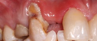
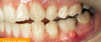
photo: earlobe swelling
Hemangioma looks like a birthmark or mole. Treatment of the tumor is surgical. The tumor is removed using cryodestruction.
Atheroma or wen is manifested by the sensation of a ball in the thickness of the lobe, which rolls inside. When you press on the tumor, a nagging pain occurs. To remove it, radio wave destruction is used.
- Why does your earlobe hurt and what to do about it?
After piercing the lobe with a gun, swelling may appear. The skin in the area of the hole turns red and becomes suppurated. A piercing in the ear requires special care, including thorough treatment of the skin with hydrogen peroxide and the application of antibacterial ointment.
If swelling of the lobe is accompanied by the appearance of a pinpoint rash on the skin, which eventually becomes covered with crusts, you should consult a dermatologist.
Why does inflammation of the middle ear occur?
As we mentioned, children are susceptible to ear diseases much more often than adults. And this is easily explained. The main reason for the development of the disease is directly related to the characteristics of the child’s auditory tube. The anatomy of the Eustachian tube is such that it is shorter and wider than the adult auditory tube. And the absence of bends allows the infection to freely penetrate from the nasopharynx into the middle ear cavity.
The causes of otitis media are:
- hypothermia or, conversely, overheating of the body;
- chronic infections in the body in children (for example, chronic inflammatory processes of the nasopharynx, from where the infection can easily get into the ear);
- adenoids;
- weak immunity;
- allergic rhinitis;
- ear damage;
- improper feeding of infants: breastfed children should not be fed lying on their back - in this position, milk can get from the nasal cavity and pharynx into the tympanic cavity and start an inflammatory process there.
Outer and inner ear: causes of inflammation
Inflammation of the outer ear occurs when trying to clean the ears with foreign objects, during which infection penetrates into them. Otitis media can occur when bacteria enter the bloodstream through wounds and damage to the skin of the ear. Therefore, parents need to ensure that the baby never picks the ear canal with a sharp object.
Some parents are overly zealous, giving their children ear hygiene and cleaning wax out of their children every day, which is fundamentally wrong. Sulfur is a natural barrier against pathogens, so such excessive cleanliness opens the way for bacteria to distant areas of the ear.
The appearance of otitis externa can be caused by water that contains pathogens getting into the ear, for example, while swimming in polluted waters.
Labyrinthitis or internal otitis in acute form manifests itself in the absence of timely treatment for the acute course of otitis media. The infection can enter the inner ear through the membranes of the brain (with meningitis) or through the bloodstream if pathogens are already present in the body. Children with this diagnosis need immediate help from an otolaryngologist. If proper treatment for inflammatory disease of the inner ear is not provided, the prognosis for life and health may be unfavorable.
Signs of otitis media
What symptoms of otitis media in children do parents encounter? Symptoms directly depend on the location of the inflammation.
With external inflammation, the visible part of the hearing organ becomes red and swollen, and the patient is bothered by itching. Another sign of external inflammation is pain when chewing food or swallowing. If a child has pain in the ear, it’s easy to check: lightly pull the earlobe, and the baby’s reaction will immediately make everything clear. Disease of the external ear can be focal or diffuse. With focal inflammation, boils appear, that is, point inflammation. As soon as the boil matures and purulent contents come out of it, the pain syndrome goes away. With a diffuse type of flow, the entire ear canal or some area of it becomes inflamed. The skin of the ear canal peels, itches, and sometimes blisters appear.
Friends! Timely and correct treatment will ensure you a speedy recovery!
In acute otitis media, the manifestations of the disease depend on the nature of the inflammation. When otitis media occurs in children with the catarrhal form of the disease, the child develops the following symptoms:
- acute pain that periodically radiates to the temples or jaw ("shoots" in the ear);
- increased body temperature;
- feeling of fullness in the ears;
- drowsiness, the baby becomes capricious and restless;
- Sometimes vomiting is possible.
If timely treatment of acute otitis in a child is not carried out at the initial stage, the disease will progress to the purulent stage. With it, the pain becomes more intolerable, and hearing is noticeably reduced. If there is a perforation (rupture) of the eardrum, suppuration begins from the ear.
If the treatment of the acute form of otitis was not carried out at the proper level or was started very late, the disease is highly likely to become chronic. With this disease, the symptoms are mild, the pain is tolerable. A chronic diagnosis is characterized by purulent discharge from the ear, since the eardrum does not have time to heal, ringing in the ears is characteristic, and hearing will gradually weaken.
With labyrinthitis, frequent dizziness, nausea and vomiting are observed.
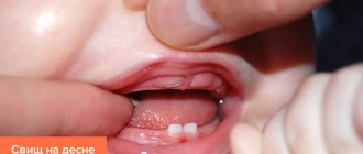
Parotid fistula: what is the danger of an additional canal in the ear
Parotid fistula is a rather unpredictable disease. In some cases, the anomaly may not bother a person throughout his life, but sometimes it makes itself felt immediately after birth and threatens with serious complications. When surgical treatment is necessary, and when surgery can be avoided, said otorhinolaryngologist at the Morozov Children’s Hospital with 25 years of experience in treating the disease, candidate of medical sciences, holder of the “Moscow Doctor” status, Alexander Mikhailovich Ivanenko.
How does the anomaly manifest itself?
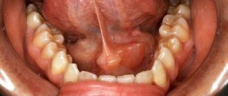
Externally, a parotid fistula looks like a barely noticeable point at the helix of the auricle. With an atypical location, the mouth can be located anywhere in the auricle, including the lobe. But this is very rare. The fistula itself is an additional passage covered with skin measuring 1.5 - 2 cm in length, which has a tortuous structure. Sometimes the fistula bifurcates or ends in a thick cyst-like extension. It can form in one ear or in two at the same time.
What are the causes of the disease?
Congenital parotid fistulas are the most common developmental anomaly of the external ear. Occurs in 15 - 43 children per 100,000, depending on the region. In 30% the disease is hereditary, in 70% it is sporadic, that is, it occurs from time to time. The appearance of a fistula is associated with abnormalities in fetal development. The defect forms at 9-11 weeks of intrauterine development. At this time, six ear tubercles are formed from the I and II gill arches, from which the structures of the auricle subsequently develop. Pathological processes during pregnancy (stress, respiratory diseases) lead to the occurrence of a defect.
Why is parotid fistula dangerous?
The fistula canal is lined with skin containing sweat and sebaceous glands, so contents of a yellow-curdled consistency accumulate and are periodically released. When a bacterial infection occurs, an inflammatory process develops that spreads to soft tissues. Huge pyogenic ulcers form on the skin, reaching 5 cm in diameter. A cosmetic defect significantly reduces the child’s quality of life. If the inflammatory process involves the cartilage of the auricle, perichondritis occurs, which leads to deformation of the auricles.
It happens that a congenital anomaly is misdiagnosed, mistaking it for other diseases: atopic dermatitis, eczema, osteomyelitis, skin tuberculosis and even systemic lupus erythematosus. Often children come to us after long but ineffective treatment.
Can parents independently detect a parotid fistula in a child?
In most cases this is possible. It is necessary to carefully examine the child and understand whether he has a small hole at the base of the ear curl or not. If there is a hole, but it looks like a small needle puncture and does not bother the child, there is no need to worry. The fistula is not dangerous and does not need to be removed. If the mouth is red, secretions are released from the hole, discomfort or pain appears - it is necessary to treat the mouth with a chlorhexidine solution and consult an otolaryngologist.
How is a congenital anomaly treated?
As a rule, patients come to us for the first time with an exacerbation of the disease. We treat the abscess and schedule a planned operation to remove the fistula. Surgical removal is performed only in the cold period, that is, not in the active phase of inflammation. Otherwise complications may arise. It takes about one and a half months to restore the skin after the inflammatory process is removed.
There are no age restrictions for the intervention. Our youngest patient was only three months old. Since birth, his fistula has become inflamed twice.
How is the operation performed?
The point of surgical treatment is to completely remove the entire fistulous tract. At the same time, the surrounding tissues cannot be damaged. A method based on the property of diaphanoscopy helps to recognize and highlight the pathological zone. My colleagues and I developed and patented the method several years ago, and we successfully apply it in practice.
During the operation, a sterile light guide catheter with a diameter of 0.75 mm is inserted through the mouth into the fistula canal. The catheter lights up and we accurately assess the length, shape and direction of the fistula, and the presence of possible branches. Then we gradually isolate the fistula from the surrounding tissues. The operation lasts from 40 minutes to an hour. This is a low-impact intervention. Previously, children spent 7-10 days in the hospital after surgery, but now they are discharged after 3-4 days. There is no need to remove sutures - we use absorbable suture material. If a child has a bilateral fistula, we remove the pathology from both sides at the same time.
We perform 20 to 30 such operations annually. Over the past years, vast experience has been accumulated in effective surgical treatment of congenital anomalies.
A baby’s ear hurts: what to do?
With kids the situation is much more complicated. An infant is not able to tell what and how it hurts, and parents can only carefully observe changes in the baby’s behavior. A sick baby becomes capricious, lethargic, and loses appetite. For no apparent reason, he begins to scream shrilly, especially during night sleep. It becomes painful for infants to suck or swallow. A sick baby constantly holds onto the sore ear or tries to lie on it to reduce pain.
Babies under one year of age are much more susceptible to inflammation of the hearing organ, because they spend a lot of time lying down, and this leads to the accumulation of mucous masses in the nasopharynx, which is an excellent environment for the proliferation of bacteria.
In some cases, vomiting and diarrhea are observed.
During treatment, infants are not prescribed ear drops, but nasal drops. Otherwise, the methods of treating the disease coincide with the treatment of preschoolers and schoolchildren.
Treatment of acute illness: where to start?
Seeing the child’s reaction to acute pain, many parents are lost and do not know what to do to alleviate the baby’s condition. At the slightest suspicion of ear inflammation, you should seek medical help, especially if you notice purulent discharge from the ear. The sooner you start treating otitis, the faster the recovery will come, and the risk of complications will be reduced to zero.
Only an ENT doctor should treat otitis media! If for some reason it is not possible to immediately consult a doctor for treatment (for example, a sharp pain occurred at night), you need to numb the ear. For acute pain, children are given medications based on paracetamol or ibuprofen (for example, Panadol or Nurofen). And in the morning you need to go to the clinic.
At the appointment, the ENT specialist will examine the child using an otoscope or a special ear specula, determine the location of the inflammatory process, its nature (the baby is suffering from an acute or chronic disease) and give recommendations for treatment.
You should not treat otitis media yourself! At home, in addition to taking painkillers, you need to carefully blow the patient’s nose, and remove mucus from the baby with a special aspirator. This should be the end of your own treatment.
Some parents mistakenly, without consulting a doctor, try to alleviate the patient’s condition and cure acute otitis in their child with ear drops. But if the eardrum has burst, using, for example, alcohol drops is not only undesirable, but also dangerous!
Treatment and prevention of atheroma
Treatment of atheroma is predominantly surgical, with removal of the cavity along with its contents. To do this, a small skin incision is made on the body, through which access to the atheroma is provided. After removing the contents, the cavity is washed, and if necessary, a turunda is inserted for several days to facilitate the release of blood, pus and fat.
The operation is performed not in a hospital setting, but on an outpatient basis. Local anesthesia is used for pain relief. For small atheromas, sutures are not applied, since the skin incision heals on its own after 5-6 days. For large atheromas, cosmetic sutures are applied, which are regularly processed after 1-2 days. The operation itself is painless for the patient, but the danger is that without eliminating the causes that caused the appearance of atheroma, there is a high probability of relapses (almost 50% of all cases). That is why it is so important not only to eliminate the cause of atheroma, but also to take measures to prevent the disease. These include personal hygiene measures and early consultation with a doctor. It is not recommended to treat atheroma on your own.