Tooth extraction is carried out plannedly, as well as for emergency indications.
The main indications for planned tooth extraction: complete or severe destruction of the tooth crown and the inability to use it for prosthetics, chronic periodontitis that is not amenable to conservative treatment, grade III tooth mobility in periodontal diseases, and others.
Urgent tooth extraction is indicated for purulent periodontitis, acute osteomyelitis, periostitis, sinusitis, lymphadenitis (if the source is a diseased tooth), fracture of the tooth crown exposing the pulp and some other diseases and conditions.
Removing various teeth has some of its own characteristics, for example, removing teeth with a crown is somewhat simpler than removing tooth roots. The extraction technique for the teeth of the upper and lower jaws, incisors and molars is also slightly different.
How is upper jaw teeth removed?
Before the operation, an examination is carried out, if necessary, radiography, consultations with other specialists. Typically, tooth extraction surgery is performed on an outpatient basis. Local anesthesia is most often used. Before the operation, the mucous membrane and teeth are cleaned to prevent infection of the socket. For tooth extraction, forceps and elevators are used, and in some cases, the tooth root is cut out with a bur. Special forceps are used to remove the wisdom tooth of the upper jaw.
Removing the upper front teeth is usually simple. Certain difficulties arise when removing the upper canine, since it has a long, massive root, the apex of which is very often curved, as well as when removing molars, since they have three roots.
When removing the upper teeth, the patient sits in a chair with a slightly reclined back and headrest. The dentist is positioned to the right and in front of the patient.
Stages of tooth extraction:
- Separation of the gums from the edge of the alveolus and the circular ligament from the neck of the tooth (with a raspatory or a trowel);
- Applying and advancing forceps under the gums, fixing them;
- Rocking or rotation of the tooth, during which the periodontal fibers that connect the tooth root to the walls of the socket are torn;
- Extracting a tooth from the socket;
- Inspection of the surgical site, extraction of small pieces of bone and tooth roots.
The success of the operation does not depend on the physical strength of the dentist, but on the correct execution of all stages of the procedure. Minor bleeding from the hole stops after 2-5 minutes, the hole is filled with a blood clot, which protects the wound from infection. To avoid damaging the clot and causing bleeding, the patient is advised not to rinse the mouth or eat food for several hours. On the day of surgery, you should not consume hot food or drinks, or take any thermal procedures; it is recommended to refrain from heavy physical activity. If all recommendations are followed, the tooth healing process will proceed quickly.
Premolar Removal For Orthodontics
Bite correction with tooth extraction is quite widespread in our daily practice. Such a “verdict” has not surprised or frightened anyone for a long time. And doctors are used to it: if anything happens, they can safely write out a referral to a surgeon for removal. And the patients resigned themselves: if it’s necessary, then it’s necessary. Where to go?
Article based on materials from Dr. Filatov R.V.
And indeed, in some cases it is impossible to do without removal. But, often, when deciding which teeth are best to remove from a given patient, many mistakes are made. Which can lead to very, very sad consequences.
Oddly enough, behind the mistakes when choosing which teeth are to be “liquidated” is a banal ignorance of anatomy and physiology. Or, no less banal, the unwillingness to make up for this lack of knowledge.
What teeth are most often removed during orthodontic treatment (for example, with braces)? That's right - premolars. "Fours" and "fives". What motivates such a choice of “victim”? And what evidence do the doctors justify?
For crowding of teeth in the frontal region.
After all, the premolars are located close to the epicenter of this crowding. Next to crooked incisors or next to fangs located outside the dentition (the upper fangs usually “hang” on top, and the lower ones, on the contrary, are located below). And when “straightening” this crowding, and even more so to return the fangs to their rightful place, the uneven teeth, straightening, take advantage of the space vacated nearby by the removed “four”. And there is no need to “drive” them all over the jaw. Everything is fixed relatively quickly and efficiently.
In the treatment of distal occlusion.
When the upper incisors protrude strongly forward. And when it is necessary to eliminate the sagittal gap. The same “fours” are located close to the canines, the distalization of which in the treatment of distal occlusion with the removal of premolars presents a certain difficulty. And here everything seems to work out optimally and rationally: the canine is in place of the removed “four”, and then all the front teeth are in place of the fang. Here you seem to have contact, bite, and happiness with health...
The removal of premolars during orthodontic treatment is also justified by the fact that they allegedly have much less functional value than the same molars. Well, it’s clear that removing canines and incisors is not comme il faut at all (although sometimes they do this). If you remove the “four”, the “five” still remains there. And she's almost the same. It doesn't seem to be a big deal. Function and aesthetics seem to have not been lost. The jaw (and the body) should not seem to notice the “loss of a fighter.” And the “hole” from the “four” is deeper and not so noticeable.
The vast majority of colleagues think so. Is this really so?
At all times, there have been serious debates between various orthodontic schools on the issue of “removing or not removing” teeth during treatment. Arguments, facts, and evidence were presented on both sides. And, I repeat, there were, are and will be indications for orthodontic treatment with tooth extraction. Always. That's not the question. The question sounds a little different: which teeth are better (more rational, safer) to remove during orthodontic treatment (with braces, in particular).
Who came up with the idea that it was the “fours” and “fives” that needed to be removed?
History is silent about this. If my memory serves me correctly - Charles Tweed.. But the fact that we were taught to easily part with premolars (not our own, of course... patients) is a fact. As historical as it is medical.
Well, well, Tweed (or whoever else...) came up with the idea of parting with teeth easily. It’s clear that I didn’t come up with the idea out of a desire to harm anyone. And from the best, humane intentions. But time flows. New facts have been obtained, new discoveries have been made, and extensive experience in using this technology has been accumulated. And therefore, as they say in jurisprudence, due to newly discovered circumstances, the question of the advisability of removing premolars requires a thorough review.
To a greater extent, this concerns the question of the advisability of removing specifically the upper premolars. Because Due to the anatomical and topographical features of their location, it is the removal of the upper “fours” and “fives” that can come back to haunt the patient with big troubles. Whereas the removal of lower premolars does not lead to such negative and destructive consequences. Due to the fact that the lower jaw, unlike the upper jaw, has a different anatomy and topography.
What are the consequences of removing premolars?
In particular, removal of upper premolars. Let's look at this issue from an anatomical and physiological aspect. And on specific clinical indications for treatment with the removal of premolars, which we were taught at one time.
First, let's remember the anatomy of the upper jaw. How is it generally structured?
In front there are two incisal bones. Pairs. Posterior to them are paired maxillaries. Next (in depth) are two palatines. Also a pair: right and left. Which, in turn, are already connected not to anything, but to the sphenoid (otherwise the main) bone. One of the bones at the base of the skull. Base of the skull! This bone is not called the main bone for nothing. It, along with the occipital bone, plays a huge role in the implementation of the so-called. the craniosacral mechanism (CSM) or, in other words, the primary respiratory mechanism (PRM), which ensures the normal function of the central nervous system and is expressed, among other things, in the rhythmic movements (microoscillations) of the skull bones.
By the way, in order for these micromovements (flexion and extension) to occur, the sutures that connect the cranial bones (and there are more than twenty of them in the skull) should not be blocked. Otherwise, of course, there will be no micro-movement. Moreover, the movement is symmetrical.
When do we most often delete “fours”? Right. When we (or rather teeth) don’t have enough space on the jaw.
And the teeth are “in a bunch.” And instead of expanding the “living space” (developing the jaw), we reduce the number of teeth. Thus perpetuating the jaw deformation.
The reason there is not enough space for the teeth is that those six bones of the maxillary complex (palatine, maxillary and incisive) are in compression, in other words, “flattened”, welded to each other. And the stitches on the upper jaw are blocked. No micro-movement. And instead of unblocking the stitches, we remove the teeth. And then what? That's right - we close the gaps from the extracted teeth.
Now pay attention to where the premolars are located relative to the bones of the upper jaw. And what happens when a premolar (usually a “four”) is removed and we try to distalize en mass the entire frontal group of teeth (incisors and canines)? It only seems to us that by closing the gaps we only move the teeth: first the canines in place of the removed premolars, and then a group of incisors.
But in fact, we move, among other things, the incisal bones towards the maxillary bones, thereby exacerbating the block in the sutures between them and “fixing” the deformity. Under the influence of the distalized incisor bones, the maxillary bones also move medially (towards the palatine bones). And under the influence of the maxillary - palatine. This is how the “tale about the turnip” turns out.
What do the palatine bones come into contact with? Where are they moving? Right. Towards the sphenoid bone, also causing displacement and compression in this area. And the sphenoid bone is, for a second, one of the bones at the base of the skull. And it is not surprising that after treatment with the removal of premolars, the patient may experience (and very often does) a completely non-dental clinic. But rather neurological. Or even psychoneurological. Headaches, neck pain, myofascial syndrome, tinnitus, blurred vision, hearing, panic attacks. What did you want? This is a skull. And any ill-considered intervention in its structures is fraught.
I'm not even talking about the deterioration of facial aesthetics: sunken lips, cheeks, protruding cheekbones, concave profile... We in no way associate these manifestations with treatment by an orthodontist. But in fact, they can be a direct or indirect consequence of treatment with the removal of premolars. Everyone now loves to talk and talk about psychosomatics.
About its cause-and-effect relationship with many, many diseases. This is a newfangled interpretation of the old common expression that “all diseases come from nerves.” Well, I can’t say for all diseases (I specialize more in teeth and jaws). But the fact that cranial (cranial) distortions constitute precisely the somatic component of psychosomatics is a fact. Literally, a medical fact. And by exacerbating these distortions with ill-conceived and unpredictable interference in the anatomy of the skull (especially by removing and moving teeth), are we often aggravating the patient’s ill health?
When else do we like to remove premolars? Remember? Yes for distal occlusion. When the upper teeth are in protrusion.
And instead of bothering to find the cause of protruding teeth at the skeletal (jaw size problem) or cranial (jaw position problem) levels (and the reason most often lies there) - we simply remove the upper teeth (usually premolars), close the gaps, trying to dock upper teeth with lower teeth in the sagittal direction. In essence, we are trying to solve “non-dental” problems at the expense of teeth (reducing their number). And we often get the problems described above. And sometimes we don’t even think that the treatment is inadequate and does not correspond to the cause. Moreover, we are aggravating this reason.
By the way, when treating distal occlusion by removing premolars, there is another mechanism for causing troubles and complications. It is no secret that most often the cause of distal occlusion is a more posterior than normal position of the lower jaw. With all the consequences... And accompanying... Deep bite. Beveled profile. Reduced height of the lower third of the face. The TMJ (temporomandibular joint) is in compression, because the head of the condyle is displaced distally (backward). What contributes to the development of degenerative changes in the TMJ.
And TMJ (or rather its dysfunction) is in itself a wide field for various kinds of non-dental clinical manifestations. What is there... For dysfunctions of the temporomandibular joints (TMD), the clinic is often more psychoneurological than dental. And there is a reason for this: compression of the joint elements, muscle spasm, which always accompanies distal occlusion and TMD, lead to improper redistribution of blood pressure in the skull, tension of the dura mater, and irritation of nerve endings. This can also cause headaches, tinnitus, and panic attacks.
But this will be a separate conversation. It's a very serious topic.
And all because the entire lower jaw, I repeat, is positioned posteriorly from the norm. And very often this incorrect posterior position is caused (and accompanied) by a distally located upper jaw. Or underdevelopment of the upper jaw in that same premaxillary zone, and therefore its (upper jaw) shortening. And instead of bringing the lower jaw forward, decompressing the TMJ, and unloading the joint to relieve the symptoms of dysfunction (and often this is pain!), we are trying to solve this problem at the level of the teeth.
And how do we decide? By removing. And by removing premolars, we get all of the above problems.
In addition, by reducing the length of the already short upper jaw, we simply “perpetuate” the distal position of the lower jaw. And thus we simply “bury” the temporomandibular joint. And we play into the hands of the further development and consolidation, already at the skeletal and dental levels, of that same dysfunction with all its symptoms. Such a vicious and vicious circle...
Regarding the small value of premolars in a functional aspect... There is also something to argue about. Which teeth make the first contact when closing the mouth? Which teeth perform the chewing movement first? Yes, exactly premolars. And these are very functionally important teeth. And their location in the jaw is a very sensitive area for all kinds of skeletal and cranial distortions.
CONCLUSION:
There are still indications for treatment with the removal of premolars. But! They are so narrow, so specific and so rare that we won't even discuss them. These indications can be classified as exceptions. And exceptions, as we know, only confirm the rules. Therefore, treatment with the removal of premolars cannot in any way be a common, routine, widespread and everyday practice. Orthodontists need to more carefully analyze the consequences of such treatment. And diagnose patients more thoughtfully. And then the number of patients with various kinds of pain syndromes, distorted faces, and simply patients dissatisfied with the treatment you provided, will noticeably decrease.
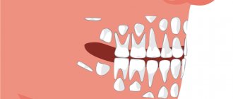
Possible complications after tooth extraction
Complications can appear during surgery, as well as after it, and can be local or general. General: fainting, rarely – shock. Their cause is the patient’s psycho-emotional stress, sometimes pain due to poor-quality anesthesia.
Local complications:
- Fracture of the tooth being removed (crown or root);
- Dislocation or fracture of a nearby tooth;
- Trauma to the soft tissues of the oral cavity;
- Dislocation or fracture of the jaw;
- Perforation of the bottom of the paranasal sinus;
- Pushing the tooth root into the maxillary sinus;
- Bleeding;
- Alveolitis.
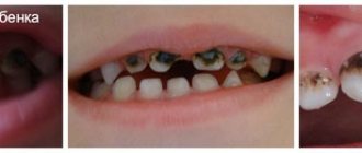
What are the dangers of premature removal of baby teeth?
Speech disturbances, inhibited bone growth and unnecessary pain are just some of the unpleasant consequences of not properly caring for baby teeth. Some parents mistakenly believe that there is no need to worry about baby teeth if they fall out anyway. Premature loss of primary teeth, that is, 3-4 years before the date of their physiological loss, in adulthood leads to unpleasant consequences - crowding or various types of malocclusion.
Baby teeth have a specific structure and are very susceptible to caries. Their premature loss is most often caused by advanced caries or mechanical injuries. If primary teeth begin to deteriorate, inflammation of the pulp quickly occurs, and then inflammation easily occurs with complications around the periapical tissues or abscesses. In such cases, it is often impossible to perform root canal treatment. All that remains is to remove the baby tooth. The whole process of pain and treatment exposes the baby to many troubles, and it is worth protecting him from this.
Indications for removal
Healthy, well-positioned third molars with good oral access for possible treatment do not need to be extracted. There are clear indications for such an operation, and if they exist, it should be removed without waiting for complications:
- Deep caries of the wisdom tooth, as well as lack of normal access for full treatment.
- Chronic inflammatory processes caused by third molars, including in cases where the wisdom tooth caused inflammation of the gums.
- Chronic injuries to the oral mucosa due to incorrectly positioned “figure eight”.
- Impacted or dystopic teeth.
- The need for orthodontic treatment of bite defects if the doctor believes that the “eights” will prevent the dentition from occupying the correct position.
In all of these situations, it is recommended to remove the “eights” without delay.
Why are wisdom teeth needed?
Why does our body need third molars at all? In fact, these are rudiments that not all people today erupt - approximately a third of the entire population of the planet does not even have the rudiments of third molars, although a few thousand years ago absolutely all adults grew wisdom teeth. They practically do not perform the chewing function, since the rest of the healthy teeth usually cope with it.
Today, “eights” are needed only as a “backup option” in case the first and second molars fall out or are severely damaged, and also as a factor preventing the loosening of neighboring teeth. They can also serve as a support for prosthetics - but, again, in all these cases we are talking about healthy and correctly erupted “eights”. In the same case, when a person has rotten wisdom teeth in his mouth, they will not be able to perform any of the indicated functions and it is better to remove them so as not to expose the rest of the dentition to the risk of infection.
Diagnosis and treatment of maxillary second molars with two palatal roots
In addition to ideal knowledge of the morphology of the root system, every doctor during endodontic treatment must also know the prevalence of individual aberrations deviating from normal anatomy in order to identify all changes and features of the root structure before treatment begins.
In clinical practice, conventional radiography and an operating microscope are, in principle, sufficient for an objective assessment of the anatomy of the root canal, but it is quite difficult to achieve any more rigorous detail using these methods. It is for this purpose that endodontics uses cone beam computed tomography (CBCT), which is simply a critically necessary diagnostic tool that allows for a comprehensive assessment of all the features of root morphology (most often the molars of the upper jaw need this). One of the most unusual and rare forms of anatomical aberration appears to be the case of a maxillary second molar with two separated palatal roots. This article presents a clinical case of treatment of a patient with the above-mentioned features of the root system. Clinical case A
26-year-old patient sought help at the Department of Therapeutic Dentistry and Endodontics of the University of Belgrade with the following symptoms, which had bothered him for several weeks:
- spontaneous attacks of pain in the area of the left maxillary molar;
- moderate sensation of pain when eating solid food.
From the anamnesis it was also possible to find out that:
- no other symptoms were observed, there was no irradiation of pain;
- this tooth underwent endodontic treatment several years ago;
- two teeth on the offending side of the upper jaw were removed at least ten years ago.
During the clinical study it was possible to establish that:
- on the left side of the upper jaw there was only a second molar (tooth 27) with a huge amalgam restoration;
- the patient showed moderate sensitivity to vertical percussion of the buccal tubercle and a painful reaction to percussion of the mesiopalatal tubercle;
- no sensitivity to palpation of the vestibular or palatal sides was detected near the disturbing tooth;
- tests for hot and cold stimuli, as well as electroodontometry showed negative results;
- No signs of pathological tooth mobility were found.
A diagnostic periapical radiograph revealed:
- partially obturated palatal and mesiobuccal root canals and unobturated distal buccal root canal;
- an area of slight radiolucency around the apex of the palatine root; lack of a distinctive border with the surrounding bony structure of the maxillary sinus.
The need for repeated endodontic treatment was explained to the patient, to which he immediately agreed, after which a plan for subsequent visits was drawn up.
Treatment procedure
The old amalgam restoration and phosphate cement liner were completely removed and the cavity walls were further prepared to provide clear visibility and straightforward access to all root canal orifices. The openings of the palatal and mesiobuccal root canals were obturated with phosphate-iodine cement material and gutta-percha pins. Approximately 3 mm distal to the orifice of the obturated palatal root canal, an oval foramen with perforation was found. Further assessment of the pulp chamber was carried out using magnifying loupes (4.5X) and an Endodontic Probe Orifice Opener (DENTSPLY Maillefer). Using a probe and a size 10 K-file, a second palatal canal (distal palatal canal) was identified in the area of the foramen ovale. The orifice of the distobuccal root canal was hidden under brown deposits of tertiary dentin and was located approximately 2 mm distal to the obturated orifice of the mesiobuccal canal and approximately 2 mm more buccal to the orifice of the distalopalatine canal. The orifice of the distobuccal canal was slightly widened with an instrument to make it easy to identify later. The second mesiobuccal root canal was not found even under the control of loupes after applying a decalcifying solution (17% EDTA).
After consultations and obtaining the patient’s consent, it was decided to carry out the entire procedure in two stages. First, using rotating nickel-titanium files, ProTapers D1, D2 and D3 (DENTSPLY Maillefer) and manual H-files (DENTSPLY Maillefer), root fillings were removed from the mesiobuccal and mesiopalatal root canals. Further instrumental processing of these canals was carried out with reciprocating movements using WaveOne files (DENTSPLY Maillefer): the mesiopalatal canal with a tool with black markings (# 40), the mesiobuccal canal with a red marking (# 25). The working length was determined and checked several times during the treatment using an electronic apex locator (RomiApex A-15, Romidan).
The distal palatal root canal was completed to approximately 1–1.5 mm from the apical foramen using size 10 and 15 K-files. The coronal part of the canal was expanded using Gates–Glidden burs sizes 3 and 2. The same procedure was performed in the distobuccal root canal. Thus, it was possible to achieve direct access and visibility of all four channels (photo 1).
Photo 1. Straight-line access to all four canals of the 27th tooth.
X-rays were taken at two different horizontal angles with K-files in all four canals, but only one image was able to differentiate all four of them (photo 2) with vague contours of the apical parts of the roots. The mesiobuccal canal was filled with calcium hydroxide, and a paper point soaked in 2% chlorhexidine solution (R4, Septodont) was left in the mesiobuccal root canal. A cotton swab soaked in chlorhexidine was left in the pulp chamber, and the cavity was closed with a temporary filling material. After two weeks, the distal-palatal and distal-buccal root canals were carefully prepared using the same WaveOne technique as for the MP and MB root canals: the distal-palatal canal with a black-marked instrument (#40) and the distal-buccal with a red (#25). The working length was determined and checked in the same way.
Throughout the endodontic treatment, solutions of 2.2% sodium hypochlorite and 10% citric acid were used as irrigants in all four canals. Each of the four canals was finally irrigated with 40 ml of 2.2% NaOCl, dried and obturated with Acroseal (Septodont) with one gutta-percha point of appropriate taper (DENTSPLY Maillefer; Figure 3).
Photo 2. Identification of all four canals of tooth 27 on a targeted radiograph.
Photo 3. Obturated orifices of the root canals of the 27th tooth.
The results of post-treatment intraoral retro-alveolar radiography were of relatively poor quality due to overlap and interference of the lower zygomatic arch and adjacent bony structures, making it extremely difficult to objectively assess the condition of the periapical tissues (Figure 4). With the patient's consent, a CBCT scan was performed, which made it possible not only to verify the result of the treatment, but also to document an extremely rare clinical case. With the formation of a small field of view (50 x 50 mm) and using the SCANORA 3Dx device (Soredex), treatment results were recorded immediately after treatment and at a six-month follow-up period. The edited images (OnDemand3D, Cybermed) clearly visualized the two separate palatal roots, as well as their position in relation to the two buccal roots and adjacent anatomical structures, but most importantly, it was possible to objectively assess the quality of obturation of all four root canals (photos 5-9).
Photo 4. Intraoral radiography of the 27th tooth did not provide objective information regarding the quality of treatment.
Photo 5. Axial cut at the mid-length of the roots.
Photo 6. Axial section in the apical region.
Photo 7. Sagittal section of the 27th tooth.
Photo 8. Detailed view of tooth 27 after treatment
Photo 9. Volumetric rendering and multiplanar reformats of tooth 27 after treatment.
Conclusions and Key Points
Careful evaluation of the internal anatomy of the pulp chamber is critical to identifying all root canals. An upper second molar with two separate palatal roots is a rare anatomical aberration, and to our knowledge such a clinical case is encountered only once every ten years. CBCT images provide more accurate and reliable information about roots and root system morphology compared to conventional radiographs. In addition, the use of CBCT images provides more predictable and successful endodontic treatment results.
Author: Katarina Beljic-Ivanovic, Assistant Profesor, Serbia
Treatment of baby teeth by a doctor
Just like adults, children's baby teeth are susceptible to caries. The treatment in the clinic is similar to the treatment of permanent teeth, with the difference that you can use special plastic fillings for baby teeth in white or many other colors. In addition to the temperature, premature removal of a baby tooth can distort the growth of the upper or lower jaw, cause insufficient space in the arch for permanent teeth, or damage their buds. This is why it is so important that the baby tooth remains in the mouth until natural tooth replacement occurs.
As permanent teeth erupt, baby teeth tend to move around a lot. This may be accompanied by purulent inflammatory processes. Then it is better to remove the baby tooth. When the first permanent molars appear, it is worth considering dental fillings. The procedure consists of fixing them with a special varnish that protects them from rotting. The treatment is completely non-invasive and painless. The optimal time for placing fillings is about 6 years.