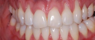
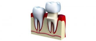
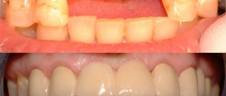
Problem: a patient approached the orthopedic dentist at the Family Dental Clinic with numerous complaints. One of the complaints was the unsatisfactory condition of the crowns (shown by arrows in the photo), soreness of the gums in the area of the crowns. After removing the old crowns, inflammation of the gums and caries on the teeth, below the gum level, were discovered. An X-ray of the teeth showed that there were poorly treated canals. To carry out high-quality prosthetics, it was necessary to lengthen the crowns of the teeth, treat root canals, cure dental caries and strengthen the roots.
Solution: complex treatment carried out by Dial-Dent specialists, a periodontal surgeon, an endodontist and an orthopedic dentist, included surgical lengthening of tooth crowns, gum treatment, treatment of tooth canals under a microscope, treatment of caries, dental prosthetics with ceramics.
Contraindications
Lengthening is not suitable in some cases, so dentists do not recommend surgery in the presence of the following conditions:
- the tooth has a short root;
- the biological width of the dental element is not enough;
- in cases where the expended efforts and resources are not justified and it is easier to carry out prosthetics;
- there is not enough space between the tooth and the antagonist;
- if the appearance of the tooth or adjacent elements deteriorates.
Professional dentists determine the feasibility in each case individually.
The perfect smile of Hollywood stars
Dental services in America are noticeably expensive. For this reason, more aesthetic and image prosthetics are carried out there, and medical procedures often remain in the shadows. The new teeth of famous personalities are often displayed like an expensive car. Unfortunately, the Hollywood smile is not for everyone. Perhaps you have noticed how comical teeth sometimes look in some photos.
The effect of “artificial dentures” is not at all what modern aesthetic dentistry strives for. Your smile should be natural and healthy, highlighting your individuality. A harmonious, attractive smile, what is it like according to the majority of our compatriots?
Removal methods
Dentists offer four ways to lengthen the crown:
1. Surgical. During surgery, part of the gum and/or bone is removed. It is carried out in the form of gingivoplasty, gingivectomy and bone resection.
2. Therapeutic. Composite extension in the incisal area. It is effective for small chips and chips.
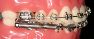
3. Orthodontic. The tooth is stretched using braces. The method is allowed if there is free space between the teeth and is recommended when lengthening one tooth that differs in length from the rest.
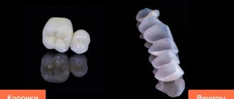
4. Orthopedic. Extension is performed using veneers or crowns. The tooth is lengthened due to the crown part without changing the gingival zone.
Surgical Crown Lengthening for Distal Restorations
Today, more and more often, in distal restorations, we are seeing the technique of raising the deep subgingival margin (Deep Margin Elevation). It is performed in clinical situations where it will be necessary to perform bone correction surgery and surgical crown lengthening. We emphasize that the procedure for raising the deep subgingival margin, recently introduced by Professor Pascal Magne, Dr. Didier Dietsky and Dr. Roberto Sprafico, is used in borderline clinical situations where rubber dam isolation is difficult but still possible; in cases where it is necessary to facilitate impression taking and subsequent isolation for bonding, and, importantly, in those clinical situations where the biological width of the periodontium is still present.
Author: Giuseppe Marchetti
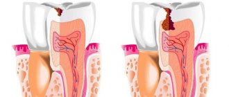
The concept of biological width, postulated by Gargiulo et al in 1961, is a fundamental requirement when performing restorations and prosthetics. The biological width must be observed both when performing restorations and prosthetics, otherwise there is a high risk of creating iatrogenic periodontal pockets.
In this article we want to demonstrate the technique using simple and easily repeatable illustrations without performing DMA. The procedure consists of the following steps. First, the carious cavity is prepared to prepare the cervical edge, which should no longer move apically. Then - an operation to place the bone crest 3 mm below the already prepared cervical edge. And finally, the restoration itself. Carrying out surgery before preparing the cervical edge is a big mistake, because... often results in the need to perform DMA.
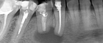
Photo 1 – Bitewing before treatment. Large carious cavity on the distal surface 1.6.
Photo 2 – In the oral cavity.
Photo 3 – After preparation, the need for surgical lengthening of the crown becomes obvious.
Photo 4 – Preparation of the carious cavity, distal cervical edge and surgical lengthening.
Photo 5 – It is necessary that the bone crest is 3 mm below the cervical part of the tooth crown.
Photo 6 – Suturing.
Photo 7 – Insulation using rubber dam. Note the good isolation obtained from the surgical procedure.
Figure 8 – The thickness of the palatal and distal cusps was less than 2 mm, so they were removed. In fact, we removed all bumps thinner than 2 mm. Advice: when preparing for a composite onlay inlay, the height must be reduced by 2 mm, for a lithium disilicate inlay - by 1 mm.
Photo 9 – Build-up, sealing of dentinal tubules, preparation for onlay inlay was carried out. Advice. Clean the dentin with glycine powder and disinfect it with chlorhexidine 2%. Then etch the enamel for 20 seconds and apply a universal self-etch adhesive to the dentin and enamel. This will allow you to avoid post-operative soreness.
Photo 10 - Only at this stage can we prepare and restore with direct restorations 1.5 and 1.7. Advice: do not do this earlier so as not to damage the restoration during the preparation of tooth 1.6 under onlay.
Photo 11 – Installation of sectional matrices and wooden wedges.
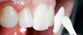
Photo 12 – Direct restorations on 1.5 and 1.7 are made in A2 shade, with brown stain added to increase the perception of depth. Finishing and polishing has been carried out. Tip 1: Use a universal self-etching bond using the selective etch technique. This will ensure the best result without any post-operative sensitivity. Tip 2: For perfect control of the working field and the absence of blood or saliva in it, finishing of restorations should be carried out in a rubber dam.
Photo 13 – Occlusal contacts are checked before impressions are taken and no corrections are required. Tip: Be sure to check the occlusal relationship and make corrections if necessary before taking impressions.
Photo 14 – Imprint. Advice: to get a result with the least distortion, it is better to make a one-stage, two-phase impression.
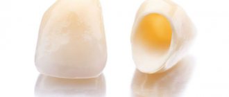
Photo 15 – Composite onlay tab on the model.
Photo 16 – After 48 hours, the insert is fitted without a rubber dam. The stitches are still not removed.
Photo 17 – Checking occlusion.
Photo 18 – Setting up the rubber dam, fitting the inlay already in the rubber dam WITHOUT bonding.
Photo 19 – Fixing, finishing and polishing.
Photo 20 - Checking the occlusion after fixing the inlay demonstrates that there is no need for correction.
Photo 21 – Final inspection of three restorations.
Photo 22 – Two weeks after surgery, good integration of restorations 1.5 1.6 1.7.
Photo 23 – Details of morphology and anatomy 1.5 1.6 1.7.
Photo 24 - Details of restorations. The tissues heal, but full recovery will occur in six months.
Photo 25 - Final X-ray. Good accuracy of the restorations was shown.
conclusions
To restore means to restore the function and aesthetics of our patients’ teeth. The restoration must be done accurately and accurately, and also last a long time. To get good results, we must follow simple rules and protocols without any flights of fancy.
If these rules are feasible, predictable and applicable, we will achieve the results we require.
After all, universal adhesives are now showing excellent results. This is not the future of adhesion, but the present, so use them without fear or doubt.
The author wishes to thank Dr. Paolo Marchetti for surgical procedures and Mr. Sebastiano Nardo for laboratory work.
The translation was performed by T. Skovorodko specifically for the OHI-S.COM website. Please, when copying material, do not forget to provide a link to the current page.
https://www.styleitaliano.org/
What should you pay attention to?
The procedure must be performed flawlessly, as even a minor mistake can cause cosmetic defects and loss of tooth stability. It is important to conduct thorough preparation, including extensive diagnostics. It includes a computed tomography scan, consultations with an orthopedist, surgeon and periodontist. This will allow you to correctly assess the condition of the jaw tissue and determine the length of the roots.
When preparing for lengthening, all points are taken into account:
- bone condition;
- aesthetics;
- condition of periodontal tissues;
- the distance between the bone crest and the gingival sulcus (should not be more than 3 mm);
- anatomy of dental roots and their relationship to the coronal part.
We create a unique, beautiful smile
A beautiful smile is not a privilege, but a requirement of our dynamic times, as well as a significant part of the image of every business person. But what makes a smile unique? First of all, it is the harmony of color, size, shape and position of the teeth. In addition, your teeth should have an optimal relationship with your facial structures.
Today, a technology has been developed for calculating the correct height of the tooth crown. It combines a mathematical calculation and a biological component, in this case the color of the iris.
Taking into account the biological characteristics of each person, beauty salon specialists will form a smile that will give the patient maximum self-confidence.
Operation stages
Most often, hygivoplasty is performed, which involves excision of the gum along the gingival contour. In some cases, part of the bone may be removed.
The operation is performed in a surgical room under local anesthesia or general anesthesia (in some cases). Preliminary sanitation is required.
The procedure itself takes place with the following steps:
- if bone resection is necessary, the flap is peeled off and an osteotomy is performed;
- a new contour is formed, which is located higher;
- stitches and a gum bandage are applied to the wound;
- For healing and recovery, antiseptic rinses are prescribed; in some cases, antibiotics are recommended. Chewing and physical activity are limited;
- removal of sutures - after 7-10 days;
- after healing, prosthetics or restoration are carried out (if temporary structures are installed, they are replaced with permanent ones after complete healing).
Final recovery occurs after 1-3 years.
Parameters of the ideal ratio of gum and tooth crown
Modern dentists have developed parameters that determine the correct relationship between gums and dental crowns, which determines the beauty of a smile. Their main provisions are as follows:
- The gingival contour on the lateral incisors should be equally spaced and located slightly lower than on the central ones;
- The alveolar process is not visible when smiling;
- The gum contour corresponds to the smile line;
- The height of the clinical crowns of the central incisors cannot be less than 11 mm.
If at least one parameter does not correspond to the above, the smile does not look attractive.
Cost of services
Surgery Price
Lengthening the crown of a tooth from 1000 rubles.
Expert of the article you are reading:
Akhmedkhanov Said Rashidovich
Dental surgeon, general dentist, implantologist, orthopedic dentist, dental therapist.
You may also be interested in:
Wisdom tooth removal Tooth extraction Removal of the dental nerve Complex tooth extraction Resection of the apex of the tooth root Removal of an impacted tooth Removal of the tooth root Removal of a dystopic tooth
Show more
What could be the consequences?
The problem most often manifests itself exclusively from an aesthetic point of view - psychological problems arise, the patient wants to correct the pathology as quickly as possible, especially if it is pronounced. That is, if the teeth and gums are healthy, then such discomfort is the only consequence of the problem.
If the short size of the crowns is caused by an abnormal structure of the teeth, problems with the gums, or abrasion of the enamel, then the consequence of this condition may be inadequate chewing of food, which will inevitably lead to problems with the gastrointestinal tract, premature aging of the face and the appearance of deep wrinkles near the mouth, and general deterioration dental conditions and even their loss.
Choosing a teeth extension technique
You can choose the optimal method of tooth extension only after consulting a dentist. The doctor will assess the degree of destruction of the dental unit, check the safety of the root and nerve, and depending on this, recommend a suitable method of restoration.
For example, veneers or lumineers are most often used to restore minor damage to incisors and canines. They hide visual defects, straighten the dentition, and return lost whiteness to the unit.
If the hard tissues of the tooth are seriously damaged, then you should resort to crowns or extensions to a pin. In this case, you can completely restore the unit and return it to functionality.
When consulting with a dentist, you need to clarify:
- The service life of the restored tooth, its resistance to wear, food coloring, and the possibility of restoration in the event of a chip. For example, some crowns can be repolished if minor defects appear; reinstallation of the microprosthesis is not required.
- The number of procedures for which it will be possible to grow a tooth. The fastest options are filling and installation of lumineers. Other technologies will require multiple visits to the clinic.
- Tissue healing rate. Some augmentation methods allow you to restore a tooth without damaging the gums. This guarantees the patient minimal discomfort after the procedure.
Different types of extensions differ in price, which must be taken into account when choosing a technique.
How is retention diagnosed?
Impacted teeth are often detected by dentists in children. They are brought to the appointment by parents who have noticed that after a baby tooth fell out, a permanent tooth did not erupt in its place. Quite often, the problem is revealed during medical examinations of children aged 11–12 years - when the eruption of permanent teeth is delayed. So, if by this time there is still a primary canine in the oral cavity, the child is prescribed an x-ray.
The ideal option for diagnosing tooth impaction is an orthopantomogram. A panoramic photograph of the jaws clearly shows all the upper and lower teeth, located both physiologically and abnormally. Some patients aged 20–30 years have baby teeth, the roots of which have not been resolved due to retention of permanent teeth. Using an overview image, the doctor determines the type of retention, assesses the complexity of the situation and develops a treatment plan.
Postoperative recommendations
After the teeth lengthening procedure, the following recommendations should be followed:
- follow all doctor's instructions to prevent infection;
- Avoid spicy, sour, solid foods that can irritate or injure tissue;
- stop smoking;
- do not overheat in the sun, in a bath or sauna, postpone taking hot baths;
- minimize heavy physical activity;
- perform oral hygiene with caution;
- treat the oral cavity with antiseptics.