A real scientific and technological revolution is currently taking place in dentistry. Cone-beam computed tomography is increasingly being introduced into the daily practice of dentists, pushing targeted and panoramic tomography into the background.
Why dentists are no longer satisfied with standard research methods, what prospects CBCT devices open for dental clinics, what advantages and disadvantages this equipment has - we will discuss all these questions in this article.
What are CBCT scanners: definition, history, operating principle
CBCT or dental tomographs are innovative devices for digital 3D tomography of the dental system, which replaced two-dimensional visualization. In just 1 scan, the device reproduces an accurate volumetric reconstruction of the maxillofacial area. Computer software allows the dentist to make various slices and sections of the three-dimensional reformat of the dental system for the fastest and most accurate diagnosis.
History of CBCT
The first CBCT scanners appeared in 1998 in Italy, they were used for diagnostics in the field of maxillofacial surgery. In their dimensions and design, the devices were more reminiscent of standard computed tomography machines. Already in 2000, Japanese scientists adopted the technology. They were the first to adapt cone-beam computer technology to the needs of dentists and developed the first dental tomograph: with a limited cylindrical scanning area of 4*4 cm and with a sitting position for the patient.
The first CBCT machines used electron-optical converters (EOCs) as image detectors. And only over time they were replaced by flat panel detectors. Unlike image intensifier tubes, flat panel sensors are more sensitive to X-rays, produce less distortion, have higher spatial resolution, and have less noise.
Today, cone beam tomography is included in dozens of different dental scanners. Modern devices differ from each other in many respects, but have a similar structure and operating principle.
Operating principle of dental tomographs
There are 3 most important elements in the CBCT device:
- X-ray tube (X-ray source)
- Planar image detector (a sensor that captures x-rays and converts them into an electrical signal)
- Movable C-shaped platform (frame connecting the detector and the X-ray tube)
During the examination, the movable platform moves around the patient's head in a circle, performing a full rotation of 360 degrees or a partial rotation of 180. As the platform moves, the X-ray tube generates radiation that passes through the object of study and is detected by the detector. The beam has the shape of a narrow cone - this shape allows you to capture a larger volume of the object in a minimum time. In total, during the study it is possible to obtain from 600 to 1200 projections of the study area. Next, the received information is processed in the system unit and reconstructed on the monitor screen in the form of a virtual three-dimensional model of the area under study.
The three-dimensional format is “cut” in layers in various planes, including axial (transverse). All received information (three-dimensional reconstruction, layers, etc.) is saved on the computer in the form of DICOM files. At any time, the doctor can return to the images to study, compare, and take the necessary measurements.
Until recently, the criteria for the use of CBCT were not precisely defined. The scope of application of dental tomographs was limited to complex cases. However, the positive experience of dentists accumulated over the years of using CBCT equipment has significantly expanded the indications for this diagnostic procedure.
Areas of application of CBCT and dental tomographs
- In therapeutic dentistry and endodontics: diagnosis of anomalies in the development of roots and root canals; caries, periodontal and periodontal diseases;
- In maxillofacial surgery and implantology: diagnosis of anomalies in the development of teeth and jaws, injuries and fractures; assessment of the prevalence of inflammatory and tumor diseases of hard tissues; planning implantation surgeries, complex tooth extractions, reconstructive interventions; diagnosis of TMJ pathology.
- In pediatric dentistry and orthodontics: diagnosis of complications of chronic pulpitis and periodontitis; anomalies in the development, formation and position of teeth and jaws.
- In otolaryngology: diagnosis and differential diagnosis of pathologies of the sinuses, nasal cavity, temporal bones.
The diagnostic capabilities of different types of dental tomographs differ. Understanding the features of modern models will help clinicians and clinic owners choose the most suitable installation for them.
Types of modern CBCT machines
Modern dental tomographs differ in scan volume (field of view or FOV), the presence of a cephalostat and patient positioning.
Field of View (FOV)
CBCT machines are classified by field of view or scanning height as follows:
- For a localized area (dentoalveolar complex): about 5 cm
- For one jaw, upper or lower: 5-10 cm
- For both jaws: 7-10 cm
- For the maxillofacial area: 10-15 cm
- For the craniofacial region: more than 15 cm
Field of view (FOV) is one of the most important parameters of a CBCT scan and depends on the size and shape of the detector, as well as the features of the collimator (a device that blocks X-rays that do not pass through the area of interest).
The larger the maximum field of view, the higher the installation cost will be. For this reason, before purchasing a device, you need to decide what shooting modes you will need so as not to overpay for equipment. For example, for implantology, an FOV of 8×8 (in some cases – 10×10) is sufficient; for otolaryngology and maxillofacial surgery – from 10×15. For devices with modular architecture, the choice of FOV can be expanded in the future.
Equipment with cephalostat
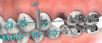
The cephalostat is a separate “arm” of the dental tomograph. It is used by orthodontists to create teleroentgenograms—images of frontal and lateral projections of the skull. The photographs allow you to study the position of the jaws relative to the skull and each other, as well as the inclination of the teeth and are mandatory when planning the installation of braces.
There are the following models on the market: with cephalostat in the basic configuration, with the possibility of retrofitting, and without the possibility of retrofitting.
Clinics where an orthodontist works require a device with a cephalostat. If orthodontic services are not provided, but are planned in the future, the medical center requires a dental tomograph with the ability to retrofit with a cephalostat.
Patient position
There are three different types of CBCT platforms, depending on the patient's position:
- Sitting position
- Standing position
- Patient supine position
Devices that scan the patient while standing are more suitable for wheelchairs and take up no more space than orthopantomographs. Sitting units are considered more comfortable, but they take up more space and make the examination of patients with disabilities more difficult. CBCT devices with the patient in the supine position occupy the largest area and volume and are not adapted for examining all patients with physical disabilities.
Advantages of CBCT scanners
CBCT scanning has a number of features that have allowed them to “squeeze out” conventional computed tomography (CT) scanners, devices for intraoral radiography and orthopantomography.
Key differences between volumetric CBCT scanning and 2D radiography methods
Higher accuracy and objectivity of images
Unlike orthopantomography and intraoral radiography, the anatomical area is scanned without loss of data, without projection distortion, overlap of anatomical structures and almost one-to-one. Thus, the 3D reconstruction obtained using a dental tomograph is an exact copy of the scanned area.
Maximum information content of the study
Cone beam tomography provides 40% more information than a conventional panoramic image.
The research allows:
- Study the position of the mandibular nerve and other vessels and nerves.
- Assess the quality and density of bone tissue, determine the height of the alveolar process (which is necessary to select the optimal solution for implantation and prosthetics).
- Determine the position of impacted teeth (teeth that are delayed in eruption) to decide on their removal.
- Visualize dental canals and assess their condition.
- Examine the condition of the temporomandibular joint (if dystopia and arthrosis are suspected).
- Investigate injuries and damage affecting the dentofacial system and maxillary sinuses.
Wide range of possibilities for working with CBCT scans using software
Volumetric reconstruction of the study area is reproduced on a personal computer. The doctor gains access immediately after the scan. He can reorient it in all three planes and precisely examine the area of interest, making CBCT slices of minimal thickness and taking the necessary measurements at a 1:1 scale. All data is saved on the hard drive in DICOM format - the doctor can return to the data at any time and continue working with it: edit or forward.
High accuracy, information content of CBCT diagnostics and ease of working with three-dimensional images allow the specialist to feel more confident in his work. The risk of medical error (missing a source of chronic infection in the area of planned implantation, touching a vessel or nerve during treatment, etc.) in this case is minimized, and the effectiveness of treatment, on the contrary, increases.
Advantages of dental tomographs compared to conventional CT scans
3D visualization of the dental system obtained using CBCT is comparable in accuracy and information content to standard computed tomography. At the same time, CBCT is superior to conventional CT in the following important parameters:
Dimensions of the equipment – more compact for dental tomographs Cost – less expensive design Software – easier to use and has a greater number of options adapted for dentistry Radiation exposure to the patient – significantly lower due to the use of innovative technologies
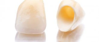
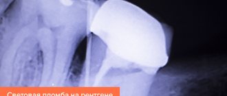
CT scan before implantation
The doctor receives reliable information about the condition of the bone tissue at the site of the planned tooth implantation. By conducting a study using a CT image, we are insured against errors; we understand exactly whether bone grafting is needed or whether an implant can be installed immediately. The image shows not only the amount of bone tissue, but also its quality. Adjacent anatomical structures are visible: the maxillary sinus, large vessels, nerve endings. Understanding the overall picture, the doctor can accurately select the implantation method and the implant itself. Without a CT scan, the dentist performs implantation at his own peril and risk!
Limitations of the use of CBCT devices
The use of CBCT in dentistry has its limitations. Thus, equipment for cone beam tomography is more expensive than orthopantomographs. In addition, the use of a dental tomograph will require new knowledge and competencies from doctors, since diagnostic information in each case must be correctly interpreted.
Another difficulty: the quality of CBCT images and, as a result, diagnostics can be affected by image artifacts, primarily metals. In modern devices, their negative impact can be reduced by fixing elements for the head and special programs, for example, the SMARF algorithm in Papaya 3D Genoray devices (South Korea).
The main disadvantage of CBCT is the inability to study the state of deep soft tissues in the study area due to limited contrast resolution. To study them, the doctor will have to supplement the diagnostic protocol with an examination on an adapted CT or MRI machine.
WHAT ARE THE ADVANTAGES OF DENTAL COMPUTED TOMOGRAPHY (CT)?
- Research speed (Gendex CB-500 tomograph scans in 8 seconds, image reconstruction in 20 seconds);
- Minimum contraindications and safety: CT is suitable for examining pregnant women, nursing mothers and young children;
- Scanning not in flat, but in three-dimensional space allows the dentist to study in detail the areas of interest from any angle, in any plane;
- Wide possibilities for diagnosing bone structures;
- The ability to record and study the image in the absence of the patient, discuss the details of the projection remotely;
- The absence of errors in image size and distortion reduces the risk of possible errors when interpreting the image.
Model range of dental tomographs. Review
At the moment, there are dozens of models of dental computed tomographs on the CBCT market. In Russia, devices made in Japan (Morita), Germany (Kavo), Finland (Planmeca) and South Korea (Vatech, Genoray) are popular. Units vary in price, patient positioning, image resolution, functionality, software capabilities, dimensions, and warranty terms.
Despite the fact that the first dental CBCT devices were developed in Japan, currently this technology is most actively developed by manufacturers from Europe (Germany, Finland) and South Korea. Dental tomographs from these manufacturers differ from their analogues in more understandable software (maximally adapted to the needs of the dentist), ease of operation, relatively compact size and affordable cost.
Table 1. Comparison of characteristics of dental tomographs KAVO, Planmeca, VATECH, Genoray
| Characteristic | KAVO (Germany) | Planmeca (Finland) | VATECH (South Korea) | Genoray (South Korea) |
| Scan time | 8-20 sec | 18-26 sec | 10-24 sec | from 7.7 sec – fastest scanning |
| Functions: CBCT, orthopantomography, TRG (cephalostat) | + | + | + | + |
| Positioning | standing and sitting | standing and sitting | standing and sitting | standing and sitting |
| Cephalostat scanning areas | 22*26 | 27*30 | 23*27 | 23*24 |
| Voxel size | 80-400 microns | 75-400 microns | voxel size (mm) 0.2/0.3 mm | 70-400 microns – highest resolution |
| Image detector | CMOS | CMOS | CMOS | CMOS |
| Current strength RT | 2-16 mA | 1-16 mA | 4-10 mA | 4-12 mA |
| RT voltage | 60-95 kV | 54-90 kV | 50-90kV | 60-90 kV |
| Focal spot | 0.5 mm | 0.5 mm | 0.5 mm | 0.5 mm |
| Shooting at low radiation dose | + | + | + | + |
South Korean Papaya 3D units produced by Genoray are in high demand among dentists. GENORAY Corporation is one of the three largest manufacturers of diagnostic X-ray equipment in Korea. The company is a full-cycle enterprise, that is, it specializes not only in the development of equipment and the production of components, but also in the assembly of medical equipment.
Distinctive features of the Papaya 3D Genoray CBCT scanner
- The maximum resolution in its class
e is 70-120 µm: for accurate diagnostics in endodontics and examination of the upper and lower dentition. - There is no need to purchase additional licenses for the software:
while with other manufacturers this needs to be done when the number of users increases. - Extended warranty of 5 years:
while the warranty for tomographs from other manufacturers does not exceed 12-24 months. - Auto positioning for all types of studies:
the function facilitates and speeds up the procedure. - SMARF algorithm:
reduces artifacts caused by foreign bodies made of metals so that they do not spoil the quality of images. - Modular architecture:
the basic device can be easily retrofitted with a cephalostat, and the design also allows for expansion of the scanning area - without replacing the device. - Maximum comfort for patients:
during the procedure, the patient is face to face with the doctor, which facilitates interaction with the patient and speeds up the examination. You can also set a voice notification about the beginning and end of the procedure. - Ability to examine patients with disabilities,
including people in wheelchairs.
Children's Study
There are cases where it is necessary to conduct this study for young children. Of course, a child’s body is more sensitive to radiation, but if there are serious indications, diagnosis should not be abandoned. If the child does not have an absolute contraindication to the study, then this method can be used even for babies in their first year of life.
Such restrictions include:
- birth injury;
- congenital anomalies;
- allergic reactions to drugs used to give anesthesia;
- heart disease.
Before children are examined, they should not be fed for 2.5 hours before the intended procedure, otherwise aspiration pneumonia may form. When at the time of the study the child is already 4 years old, you should talk to him. At the same time, try to explain the procedure, be sure to emphasize that mom and dad will be nearby at all times.
For younger children, the examination is performed under anesthesia. Moreover, during the diagnosis, parents can be with the baby and wear lead aprons for protection.
Recommendations for purchasing CBCT dental tomographs
The choice of a CBCT device is made based on the needs of the clinic, software capabilities and equipment costs.
Thus, before purchasing a device, ask yourself the following questions:
- How much money are you willing to spend on a dental tomograph?
- Which specialists will operate the CBCT machine and what procedures will be performed?
- What are the dimensions of your office and are you limited in space?
- Why do I need this, and what benefits will I get from purchasing a CBCT?
Benefits of purchasing a dental tomograph
Increased income for the clinic
A CBCT device is a profitable investment for a clinic. The purchase of the device allows the clinic to expand its range of services with more popular and expensive diagnostic procedures. So, if an orthopantomogram in Moscow costs from 1,000 to 3,000 rubles, then 3D tomography costs from 2,000 to 10,000 rubles. At the same time, a CBCT device pays for itself as quickly as an orthopantomograph - due to the high price of the procedures and the large number of indications for the procedure (see above).
If the device performs 5 procedures per day (at an average price of 4,500 rubles for examining two jaws), you will pay for a mid-price dental tomograph costing 6-8 million rubles. in just 1 year. The second year of operation will bring you 8,212,500 rubles. (4500*5*365) – with the same frequency and cost of procedures and excluding expenses.
Own diagnostic center at your disposal
If you purchase a dental tomograph with a cephalostat and an orthopantomography function, then you have a full-fledged diagnostic machine at your disposal. You close the cycle of treating and diagnosing patients within your clinic. Thus, you not only attract new clients, but also eliminate the possibility of losing a patient by sending him to another dental center for diagnostic procedures.
More patient trust
CBCT images display pathological areas and anatomical features of the dental-facial system so clearly that they become understandable even to non-specialists. The patient should no longer take the doctor's word for it. For example, a doctor can clearly demonstrate to a patient the number of canals in a tooth that require filling, and thereby justify the cost of the procedure.
After canal treatment, implant installation or surgical treatment, the doctor can take an image showing the positive results of his work, which increases the patient’s confidence and motivation to continue treatment or rehabilitation procedures in your clinic.
Increasing the prestige of the clinic and increasing the professionalism of specialists
By installing the device, you will dramatically increase the status of the clinic and the level of your staff: when such equipment appears in the clinic, dentists, learning how to use it, improve their professional level.
Protection from lawsuits
During forensic medical proceedings, visual CBCT images can play an important role, namely, confirming the absence of a medical error. Moreover, all information obtained during examinations is stored in the database of the CBCT computer system: the doctor can access it at any time.
General overview
CBCT, or cone beam tomography, is a computer diagnostic method implemented using a tomograph, which allows one to obtain a three-dimensional image of the area of the jaw being examined. The technique is used in dental treatment, in preparation for surgical operations, as well as in otorhinolaryngology. As with x-rays, the examination procedure involves exposing the patient to a small dose of radiation.