Sign up for a consultation 8 (800) 550-72-25
specialist
Prices for services Get a consultation
Endoscopic operations are performed for tumors of the large intestine. For flattened or flat areas of neoplasia, the dissection method is used - blunt cutting of tissue. An infiltrate is created in the submucosal layer of the colon, due to which protrusion of the mucous membrane occurs. Then the tumor is removed. Unlike resection, endoscopic dissection uses several special knives rather than a single cutting loop. After the tumor area is raised above the submucosal layer, the entire conglomerate is removed at once.
Endoscopic surgery for tumor dissection in the submucosal layer of the large intestine is performed for flattened and flat forms of epithelial neoplasia. The affected area of the mucous membrane and part of the submucosal layer are removed, which corresponds to the rules of surgical ablastics - excision of the tumor within healthy tissue.
Find out prices for paid services
| Service | Price |
| Initial consultation | 2,100 rub. |
| Repeated consultation appointment | 1,400 rub. |
| Endosonography | 10,900 rub. |
| Endoscopic mucosal resection | 14,600 rub. |
| Endoscopic dissection in the submucosal layer | 55,000 rub. |
| Endoscopic ligation of the polyp base | 9,100 rub. |
| Endoscopic polypectomy up to 2 polyps | 6,000 rub. |
| Endoscopic polypectomy of more than 2 polyps | 7,400 rub. |
| Removal of polyp of the anal canal and rectum | 6,400 rub. |
| Endoscopic biopsy from the colon in 1 (one) zone | 540 rub. |
| Endoscopic stenting of the area of tumor stenosis | RUB 82,500 |
| Histological examination 1st category. difficulties | 1,400 rub. |
| Histological examination 2 cat. difficulties | 2,700 rub. |
| Histological examination 3 cat. difficulties | 4,000 rub. |
| Histological examination 4 cat. difficulties | RUB 5,150 |
How is endoscopic dissection performed in the submucosal layer?
Under intravenous anesthesia, as with a regular colonoscopy, an endoscope is inserted through the rectum to the site of the tumor. Then, using a thermocoagulator, the pathological area is limited from healthy tissue. Cauterization of vessels along the tumor borders prevents the development of bleeding during endoscopic dissection. Then a solution of sodium salt of hyaluronic acid is injected into the submucosal layer, due to which the damaged area is peeled off and raised. After this, using several knives, the tumor area within the boundaries, designated by a thermocoagulator, is separated. Endoscopic dissection in the submucosal layer ends with the removal of a single block of tumor cells. The removed area must be sent for histological examination.
The time of endoscopic surgery using dissection technology in the submucosal layer takes no more than one and a half hours. The hospitalization period is 4–5 days.
ESD - Endoscopic Submucosal Dissection - Olympus Solutions for Colon and Rectum
ESD (Endoscopic Submucosal dissection) - endoscopic dissection of the submucosal layer - is a minimally invasive, highly effective method for treating pathological formations. ESD is a standard procedure for early stages of gastric cancer, such as superficial esophageal cancer and gastric cancer. However, for the colorectal region, endoscopic dissection is not a standardized technique due to some technical difficulties. To this end, Olympus has partnered with leading colorectal cancer experts in Japan to develop specific ESD tools and solutions, which are included in the latest EndoTherapy catalogue.
Most colorectal tumors are benign adenomatous lesions, and most early-stage cancers are no more than 2 cm in diameter, meaning they can be resected en bloc using the EMR technique using a surgical snare. Tumors that have spread laterally are more difficult to resect using this technique. Since most of these granular tumors are adenomatous lesions, even if large, and accurate preoperative diagnosis is possible with magnifying endoscopic examination, they can be completely removed using a well-planned EMR technique, removing the mucosa piecemeal. It is important to have a good understanding of the anatomical features of the colorectal region, as well as the characteristics of colon and rectal tumors, so as not to undergo an unnecessary ESD procedure, which may be recommended only because an accurate and qualified diagnosis of tumors cannot be performed before treatment. Since the colon is a long, hollow organ, functional disorders caused by local resection of its part, with the exception of the lower parts of the rectum, are much less problematic compared to cases of resection of the esophagus and stomach. Surgery on the lower part of the rectum can also be performed through the anus (transanal resection). Since the duration of the ESD procedure is quite long, the extent of its implementation may be controversial in case of massive lesions, since there is an alternative to performing laparoscopic colectomy. (Source: Olympus ESD. Endoscopic submucosal dissection. Technique for the colon and rectum. A manual for practitioners.)
Suggested indications for performing ESD in the colorectal area:
- Non-granular tumors spreading laterally (LST-NG);
- Type VI lesions according to the classification of mucosal relief;
- Submucosal (SM) carcinomas with limited invasion;
- Large deep-seated lesions;
- Large raised lesions;
- Lesions inside the mucous membrane, in combination with fibrous changes in the submucosal layer;
- Single local tumors localized in the area of chronic inflammation;
- Local residual early carcinomas after endoscopic resection;
Difficulties in performing an ESD procedure in the colorectal area depending on the region:
| Region | Peculiarities |
| Rectum | Retroflection required. Special treatment is required due to the large number of blood vessels. Perforation is possible when maneuvering the endoscope - caution is required! |
| Sigmoid colon | The endoscope is prone to paradoxical movements, which complicate treatment procedures. Because the lumen is narrow and curved, it is important to provide a field of view. |
| Descending colon | The limited maneuverability of the endoscope will sometimes make the procedure difficult. Particular attention should be paid to the area around the splenic flexure due to respiratory movements. |
| Transverse colon | In certain areas, the endoscope is prone to paradoxical movements. Difficult visibility in the area of the splenic flexure, the middle part of the transverse intestine and the hepatic flexure. |
| Ascending colon | A long procedure can lead to prolapse of the sigmoid colon. |
| Cecum | The ESD procedure is difficult because the intestinal wall is thin, and the knife must be directed perpendicular to the thin wall. |
Technique of ESD procedure in the colorectal area:
Careful preparation is required before starting ESD. Labeling is most often not required because the size of the lesion in the colorectal area is easily distinguishable.
- To ensure an efficient procedure, it is recommended that tissue dissection and preparation be planned in advance.
- Use local tissue injection only in areas where you plan to make an incision.
- Repeat incisions in the mucosa and preparation in the submucosal layer until the affected area is completely isolated.
- To expand the submucosal layer and increase the safety of the procedure, use local injection and change the position of the patient's body if necessary.
- Frequent hemostasis is required to ensure clear visibility.
- Inspect the bottom of the ulcer after treatment and treat, using hemostatic forceps, blood vessels from which delayed bleeding may occur.
Recommended Olympus ESD Tools:
- HookKnife (L-shaped hook)
- DualKnife
- Crocodile tongs Coagrasper
You can find a complete list of instruments for endoscopic dissection of the submucosal layer from Olympus in the section POLYPECTOMY, MUCOSA RESECTION (EMR) AND SUBMUCOUS DISSECTION (ESD) by following the link. You can buy or order Olympus EndoTherapy endoscopic instruments by calling +7 495 783 68 11 or by sending a request by email [email protected]
Where is endoscopic dissection performed in the submucosal layer for colon cancer?
Endoscopic dissection in the submucosal layer of any part of the large intestine is carried out in our department of abdominal oncology at the Research Institute of Surgery and Emergency Medicine of the St. Petersburg State Medical University named after Academician I. P. Pavlov. Our specialists will determine the conditions for conducting a minimally invasive operation and provide the necessary assistance in full.
Advantages of our clinic
- Multidisciplinary and well-equipped all departments.
- Modern diagnostic equipment.
- Reasonable prices for research.
- Highly qualified medical staff.
Intraoral submucosal fibrosis (oral submucosal fibrosis)
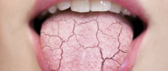
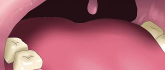
The period between the onset of tobacco use and the onset of clinical symptoms of oral submucosal fibrosis varies greatly, ranging from several months to several decades, depending on the type of betel nut consumed, the duration and frequency of the habit, individual predisposition, and other factors. Symptoms and signs of oral submucosal fibrosis occur as a result of inflammatory processes and, more importantly, fibrosis. The most common primary symptoms and signs of the disease are a burning sensation, dry mouth, paleness of the oral mucosa and the appearance of ulcers. A burning sensation usually occurs when chewing spicy food. Pallor of the oral mucosa is the result of local vascular damage due to developing fibrosis and is characterized by milky pallor of the membrane. It can be localized, diffuse or network in nature. In some cases, blanching of the mucous membrane may be accompanied by the appearance of small vesicular formations, which, when bursting, cause erosion. Patients complain that these vesicles form after eating spicy foods, suggesting the possibility of an allergic reaction to capsaicin. These symptoms are observed at all stages of oral submucosal fibrosis.
In later stages, an important feature of the disease is restriction of oral fluid due to the formation of collagen fiber bundles, which causes difficulty in chewing, speaking, swallowing and maintaining oral hygiene. The development of fibrosis in the lip area leads to their thickening and softening, making it difficult to retract and extend. Areas of fibrosis around the lips give the mouth an elliptical shape. The cheeks become thick and stiff. When the patient tries to blow a whistle or inflate a balloon, the cheeks do not change their position, as is usually the case. Depapillation of the mucosa at the tip of the tongue and along its lateral edges may be accompanied by blanching or fibrosis of the ventral mucosa. Fibrosis of the tongue and floor of the mouth prevents tongue movement. Involvement of the hard palate in this pathological process is characterized by extremely severe blanching of the mucous membrane.
Fibrosis can spread to the back of the mouth, affecting the soft palate and uvula, which shrinks or, in the most severe cases, takes on a glandular texture. Gum damage is relatively rare and is characterized by fibrosis, pallor and loss of healthy texture. In rare cases of severe damage, hearing loss may occur as a result of blocked eustachian tubes, as well as difficulty swallowing due to fibrosis of the esophageal tubes.
Submucosal layer
Submucosal layer . The proper layer of the mucous membrane, without a sharp boundary, passes into the submucosal layer (lamina submucosa), consisting of looser connective tissue. The lamina muscularis mucosae, characteristic of the mucous membrane of the digestive tract, which separates its own layer from the submucosal one, is absent in the wall of the oral cavity. In some places of the oral cavity, the submucosal layer is not expressed at all, for example, in the mucous membrane of the tongue and gums, as well as in the lateral parts of the hard palate and in the area of the palatal suture. In these places, the mucous membrane is fused with the intermuscular connective tissue or with the periosteum of the corresponding bones.
After this general characteristic of the oral mucosa, it is necessary to consider some features of the structure of its individual sections.
Lips. In the lip area, there is a gradual transition of the skin covering the outer surface of the lip into the mucous membrane of the oral cavity. The transition zone is the red border of the lips, which is found only in humans. In this area, hair and sweat glands disappear, but the sebaceous glands remain. There are more of them in the upper lip, especially in the area of the corner of the mouth. The excretory ducts of the sebaceous glands open directly on the surface of the epithelium. When these ducts are blocked
glands, their enlarged bodies become noticeable in the form of yellowish-white grains visible through the epithelium (Fig. 118).
The stratified squamous epithelium of the red border of the lips exhibits keratinization. However, the stratum corneum here is thinner than in the skin. It also has a well-defined granular layer, in the cells of which grains of keratohyalin are visible. The layer of its own, located under the epithelium, is a direct continuation of the dermis of the skin. It forms numerous papillae that penetrate deeply into the epithelium. Wide capillary loops, which are rich in the papillae, come close to the surface and are easily visible through the epithelium. This explains the red color of the lips.
The mucous membrane covering the back surface of the lips is lined with a thicker layer of epithelium, the cells of which contain a large amount of glycogen in their protoplasm. There is no keratinization here (Fig. 119).
The papillae of the lamina propria are few and rather short. Numerous small mucous and mixed salivary glands (glandulae labiales) appear in the lamina submucosa. In the thickness of the lip there are bundles of muscle fibers m. orbiciilaris oris. Intermuscular connective tissue is tightly fused with bundles of collagen fibers of the submucosal layer. This promotes smoothness of the mucous membrane of the lip and prevents the formation of folds (Fig. 120 and 121).