Periapical abscess
A periapical abscess develops as a result of infection of the root canals during acute periodontitis or in case of exacerbation of a chronic destructive process. The main causes of inflammation of the apical periodontium include injuries accompanied by rupture of the neurovascular bundle, thermal overheating of the pulp during the preparation of vital teeth before prosthetics, and toxic effects on the pulp of photopolymer materials during deep caries.
A periapical abscess can also occur as a result of infection of periodontal tissues with scabs of dentinal tubules in case of violation of the endodontic treatment protocol, as well as in the case of inadequate obturation of the canals with permanent filling material. Predisposing factors contributing to the development of periapical abscess include hypothermia and frequent infectious diseases that reduce the body's reactivity.
During the decay of the pulp, along with infiltration of the walls of the root canal by bacteria and their toxins, penetration of pathogenic microflora beyond the root apex is observed, which leads to the development of periodontal inflammation. Polymorphonuclear leukocytes are the first to rush to the pathological focus. Macrophages bind bacterial cells, activate the complement system, as a result, microcirculation is disrupted and vascular permeability increases.
The accumulation of mononuclear lymphocytes and macrophages leads to an abundant release of lysosomal enzymes that catalyze the activity of osteoclasts, which entails the destruction of the periodontium and adjacent tissues. Leukocyte infiltration promotes the formation of microabscesses, the fusion of which forms one abscess. The histological picture of a periapical abscess is represented by an area of tissue melting, along the periphery there is a shaft consisting of neutrophils.
Classification and symptoms of periapical abscess
Based on the nature of their course, periapical abscesses are divided into 2 categories:
- Periapical abscesses with fistula
. They develop as a result of a long-term inflammatory-destructive process, with granulation tissue growing into the bone marrow spaces and mucosa. It is possible to form an anastomosis with the skin, maxillary sinus, nasal cavity, and oral cavity. The disease has a chronic course. - Periapical abscesses without fistula
. They arise as a result of transcanal infection of periodontal tissues. Characterized by a pronounced clinic.
When a periapical abscess forms, patients complain of intense dull throbbing pain in the area of the causative tooth. Unpleasant sensations intensify when chewing. The configuration of the face with a periapical abscess changes due to the formation of swelling. If an abscess forms on the upper jaw, mouth opening is not impaired. In the absence of timely treatment of exacerbation of chronic periodontitis of the lower molars (wisdom teeth), the masticatory muscles are involved in the inflammatory process, resulting in contracture of the lower jaw. Patients indicate restrictions when opening the mouth, pain when swallowing.
With a periapical abscess on the oral mucosa, a painful swelling is detected in the projection of the apexes of the causative tooth. The fluctuation symptom is positive. The transition fold is smoothed. The mucous membrane is hyperemic. Vertical percussion is sharply positive. Periapical abscess is most often localized on the buccal side of the alveolar process. If the infection has entered the periodontium from the palatal canals of the second premolars or molars, an abscess is found in the palate.
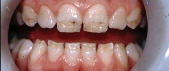
There may be a deep carious cavity or filling on the chewing surface of the causative tooth. The tooth is discolored. With a periapical abscess, the regional lymph nodes are enlarged. The general condition is disturbed. Patients complain of malaise, headache, and fever. With a periapical abscess with a fistula, the clinical picture is blurred. As a rule, the causative tooth is gray in color. Vertical percussion is painful. Lukomsky's symptom is positive. A fistula with granulations is detected on the mucosa.
Periapical pathologies: how to treat?
What is the main determining factor for a dentist when choosing an intervention algorithm during endodontic treatment: existing knowledge, experience or the results of CBCT analysis? This article describes a clinical case of a 10-year-old patient with periapical pathology, which confirms the idea that endodontically affected teeth should not always be extracted. Even in cases of roots with very curved morphology, successful treatment results can be achieved using special nickel-titanium files.
Although clinical dentistry is rich in evidence in favor of various treatment algorithms, in most cases such evidence is of very low quality. In the end, doctors rely more on their own experience. Even a simple clinical case can be treated using different methods with a fairly good prognosis. Discussion of such topics is very common on social networks. A study recently conducted at Ghent University in Belgium found that dentists are often biased and make mistakes when making necessary clinical decisions. For example, in cases of persistent and chronic asymptomatic apical lesions, the endodontist will likely opt for repeat endodontic treatment or apical surgery rather than extraction followed by implantation. Orthopedists and surgeons, on the contrary, will lean towards the latest intervention algorithm. In fact, the choice of treatment method should take into account the wishes of the patient after he has signed an informed consent that he is fully aware of all possible rehabilitation options. In this article we will discuss the best way to select a treatment algorithm in the presence of a specific apical lesion.
Endodontic treatment
Recent data from Ghent University and other epidemiological studies show that in 22% of cases, doctors choose extraction as a treatment method for periapical lesions larger than 1 cm. If the size of the pathology exceeded 1 cm, more than 50% of doctors were inclined to remove it. In principle, the choice of such intervention tactics is logical: doctors do not know the true histological picture of the lesion (whether it is a cyst or a periapical granuloma), therefore, the treatment algorithm is based on the obtained radiological data. Naïr points out that it is simply impossible to differentiate between cystic and non-cystic lesions based on radiographs alone. Similarly, during dynamic monitoring it is difficult to say whether the lesions are enlarging or shrinking after treatment.
Moreover, the limitations of 2-D radiographs must be taken into account during clinical intervention planning. Wu et al showed that targeted images often indicate a healthy apical region or extensive healing, when in fact the CBCT results show significant signs of apical periodontitis (Figures 1 and 2).
Photo 1. X-ray showing endodontically treated tooth No. 11 and cold-sensitive tooth No. 12.
Figure 2: CBCT findings from the same region of interest showing the presence of a large apical defect.
Although CBCT allows a more accurate determination of the presence and size of the apical lesion, there has not yet been enough research to support the need for CBCT scanning as a standard diagnostic method in endodontic practice. Morris et al concluded that endodontic treatment of teeth followed by direct and indirect restorations provides fairly predictable rehabilitation results, which, in turn, justifies endotherapy as the first choice.
Size matters?
In general, literature data suggest that the larger the periapical lesion, the less favorable conditions for its healing. In principle, everything is logical: the larger the pathological focus, the longer it took for it to grow. The longer a bacteriological association has developed, the more different bacteria are included in its composition, which means the more difficult it will be to cure such a pathology. Of course, a number of additional studies still need to be conducted, but in general, an increase in the focus of periapical lesions by every millimeter reduces the prognosis of successful treatment by 14 percent compared to clinical cases of endotherapy in which there are no radiological signs of periapical pathology at all. A similar trend is observed in periapical surgery: there is a negative correlation between the size of the apical lesion and the possibility of complete healing of the intervention area. During orthograde endodontic intervention, apical lesion size greater than 2 mm is categorized as a risk factor for the need for future endodontic retreatment. However, it must be remembered that the size of the formation on periapical images largely depends on their quality and positioning angle. Although it was previously believed that the larger the periapical lesion, the more likely its cystic structure, however, such conclusions are unsubstantiated without additional histological studies. Large periapical formations are characterized by a tendency to increase with the possibility of involving adjacent anatomical structures in the process. This does not mean that treatment of such lesions is impossible, but its predictability becomes much less predictable.
Clinical case: a young patient with a chronic apical abscess
A 10-year-old patient was referred for endodontic treatment of a mandibular right molar. In the area of tooth No. 46, the presence of a fistulous tract on the palatal side was noted. The patient and his parents were informed that the success rate of future endodontic intervention was reduced given the enormous size of the periapical lesion. However, they still did not want to remove the problematic tooth. After clinical examination and radiological control, a diagnosis of pulp necrosis and chronic apical abscess was made (Figure 3-4).
Photo 3. X-ray of the area of tooth No. 46 with visualization of the periapical area of the lesion.
Photo 4. Clinical appearance of the affected area.
Probing of the fistula tract confirmed its direction towards the apex. The choice of endodontic treatment was also justified by the need for corrective orthodontic and orthopedic interventions in cases of tooth extraction at such a young age. During the first visit, the tooth was isolated using a rubber dam, after which the doctor cleaned out all existing caries. To process the canals, NiTi files (HyFlex EDM) from the Swiss manufacturer COLTENE were used (photo 5). The canals were processed up to sizes 40/04. This system was used during the treatment because it is characterized by high flexibility of the instruments, which was extremely important in the patient’s existing canals with a curved morphology. In addition, only a few instruments were required to process the entire endospace. Chemical treatment of the canals was carried out using a 5.25% sodium hypochlorite solution and ultrasonic activation. After placement of an interim endodontic dressing with unsaturated calcium hydroxide, the tooth was subsequently restored using a composite restoration. To minimize the effect of microleakage, the tooth was covered with Teflon tape between visits.
Photo 5. Sequence of HyFlex EDM files.
Development of channels up to file size 60/02
During the second visit, it was noted that the patient continued to have a fistulous tract, but the swelling had almost completely subsided (Figure 6). The tooth was again isolated using a rubber dam, after which the medial canal system was treated to 50/03, and the distal canal system to 60/02. Medicinal treatment of the canal was performed with sodium hypochlorite and citric acid (40%). Since no discharge from the endospace was noted after drying, the mesial canals were obturated with bioceramics. The distal canal was filled with a plug made of MTA. The top of the tooth was covered with a composite restoration (photo 7). After three months, no fistula tract was noted, and radiographs confirmed healing of the periapical area (Figures 8-9).
Photo 6. Healing of the fistula tract.
Photo 7. Quality control of obturation.
Photo 8. X-ray after 3 months.
Photo 9. Clinical appearance of the tooth after 3 months.
At the same time, the doctor should always remember the main parameters that justify the advisability of extraction instead of endodontic treatment:
- doubts regarding the success of endotherapy on the part of the dentist who referred the patient;
- a large area of the lesion on the resulting x-ray;
- no signs of healing of the fistula after installing a calcium hydroxide bandage;
- compromised initial periodontal status;
- taking into account age-associated features of the clinical algorithm of endodontic treatment.
Is radiographic success everything?
Considering the studies mentioned in this article, the presented case shows that prejudice and subjective diagnosis should not prevent us from performing endodontic treatment even in cases of large apical lesions. With informed consent and proper motivation on the part of the patient, root canal therapy should always be the first choice of iatrogenic intervention. Although conventional 2-D imaging, which is used for early diagnosis, may hide the presence of prolonged apical periodontitis, CBCT allows us to objectify all the necessary parameters of the periapical lesion. At the same time, neither two-dimensional nor three-dimensional diagnostic methods make it possible to differentiate the differences between scar tissue in the area of the “healing” apex and the infectious process in the area of the affected apex in the absence of corresponding clinical symptoms. Based on this, success on a radiograph is not always clinical success.
Conclusion
Combining several imaging tools helps endodontists make the correct diagnosis and select the most appropriate treatment algorithm for each individual clinical case. With modern NiTi file systems, root canals with very curved morphology can be effectively treated. Ultimately, all this contributes to the fact that those teeth that were categorized as unsuccessful and planned for removal, it turns out, can still be treated if you approach the diagnostic and planning stages correctly.
Author: Dr Christophe Verbanck (Belgium)
Gum abscess - symptoms and treatment
Gum abscess is caused by pathogens of the oral microflora, which is mainly represented by the following bacteria:
- Bacteroides forsythus (in 55.6% of cases);
- Porphyromonas gingivalis (50.0%);
- Fusobacterium nucleatum (44.4%),
- A. actinomycetemcomitans (38.9%);
- Prevotella intermedia (38.9%) and others.
In addition, in most cases, periodontal pathogens enter into symbiosis with fungi of the genus Candida (44.4%), bacteria Enterobacter (38.9%), Streptococcus intermedius (13.2%), Peptostreptococcus micros (13.2%) and Staphylococcus aureus (28.5%) [4].
The pathogenesis of the disease varies depending on the location of the inflammation.
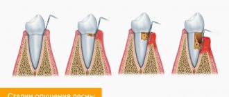
1. An abscess that arises as a result of pathological processes in periodontal tissues. Inadequate treatment of tooth root canals leads to acute inflammation or exacerbation of its chronic form in the periodontal area. As a result, the number of microorganisms in the tooth root canal and in the periodontal area increases significantly. In response to this, granulation tissue (connective tissue formed during healing) grows in the area of the root apex, replacing bone tissue with it. At the moment of exacerbation of the process, foci of purulent melting of granulation tissue appear in this area. As a result of periodic exacerbation of the inflammatory process, the periodontal lesion gradually spreads to more and more new areas of the bone, mainly towards the vestibule of the oral cavity. This leads to the appearance of vertical slit-like defects of the cortical plate and the further development of first infiltration in the gum area, and then the appearance of an abscess [5].
2. An abscess that has arisen in the area of the periodontal pocket. Periodontal gum abscess occurs as a result of several interrelated pathological processes. A large amount of supra- and subgingival dental plaque promotes the development of periodontal pathogenic bacteria, which causes a strong immune reaction. The body responds by forming an infiltrate with a large number of immune system cells. These cells produce inflammatory mediators that stimulate the destruction of bone tissue by osteoclasts (cells that remove bone tissue by dissolving mineral content and breaking down collagen). The bone tissue begins to dissolve and a periodontal pocket is formed, which, depending on the severity of periodontitis, has different depths. At the time of exacerbation of periodontitis, suppuration from the periodontal pocket becomes continuous. If the outflow is disrupted, abscess formation occurs [6].
3. An abscess that has arisen in the area of peri-implant tissue. The cause of the development of peri-implantitis is microorganisms located in the biofilm on the surface of the structure. Colonies of microorganisms affect the soft tissue surrounding the implant, causing inflammation. Further, the process develops in the same way as with periodontitis, but does not remain localized, but spreads towards the apex of the implant. In peri-implantitis, the inflammatory infiltrate passes directly to the alveolar bone, while in periodontitis it is separated from the bone by a one-millimeter layer of connective tissue. At the early stage of peri-implantitis, an abscess occurs at the level of the upper third of the implant - in the area of the peri-implant pocket. At later stages, as the bone in the area of the implant body is destroyed, it can move closer to the transitional fold of the gums[7].