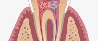
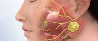
Functions of the facial nerve
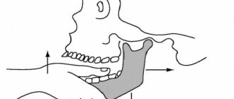
The facial nerve innervates (provides communication with the central nervous system) the facial muscles, which are responsible for the movement of facial muscles and the expression of all our emotions. There are only 2 facial nerves: right and left, each of which is responsible for the corresponding half of the face. The facial nerve has five branches: the 1st branch innervates the muscles of the upper third of the face, the 2nd and 3rd innervate the muscles of the middle third of the face, the 4th branch is responsible for the movements of the lower third of the face and the 5th branch activates the facial muscle of the neck. – m. platysma. The facial nerve exits the skull as a single trunk through the stylomastoid foramen of the pyramid of the temporal bone. Division into branches occurs in the area of the parotid salivary gland. Violation of the integrity of the nerve will lead to paralysis of the facial muscles that it innervates. Accordingly, if the nerve damage occurred in the cranial cavity, in the canal of the temporal bone and also before the branches depart, then paralysis will manifest itself on the entire half of the face corresponding to the side of the damage. If one of the branches or several branches of the facial nerve is damaged, then paralysis will occur only in those areas where the muscles that innervate these branches are located. The most severe degree of paralysis of facial muscles is damage to the facial nerve at the level of the trunk.
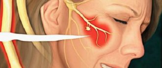
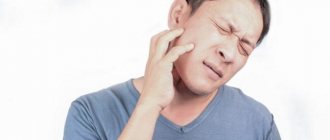
How does facial paralysis manifest? Symptoms of facial paralysis
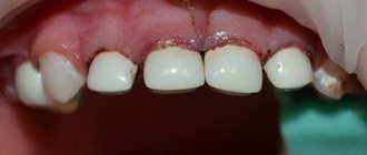
This manifests itself as follows: half of the face, or some part of it, does not express any emotions, there are no movements. A person cannot smile, be surprised, etc. At rest, asymmetry of the face is observed, and the side on which the facial nerve is damaged is lowered (the eyebrow, lower eyelid, corner of the mouth are lower, the cheek tissues sag). With facial expressions, the asymmetry intensifies. All this leads to a decrease in the social activity of people with this disease, and difficulties arise when communicating with people. But this problem is not only an aesthetically unsatisfactory appearance. There are also a number of functional disorders:
1) Impaired diction, the pronunciation of many sounds is difficult, speech is slurred, due to the lack of tone of the orbicularis oris muscle and the muscles of the buccal region; 2) Difficulty eating, especially liquids; 3) Difficulty brushing your teeth; 4) Drooling is not uncommon; 5) Complaints about the absence of tears, incomplete closure of the eyelids, which leads to the development of keratitis, conjunctivitis, which may result in the loss of the eyeball. In this case, quite often they resort to blepharorrhaphy (stitching of the eyelids), in which it is necessary to constantly use eye drops and ointments; 6) The opposite situation may also occur, in which there is constant lacrimation associated with the lack of adherence of the lower eyelid to the eyeball; 7) The appearance of difficulty in nasal breathing due to the fact that the nasal muscles do not work and a valve effect is formed.
All of the above symptoms significantly reduce the quality of life of a person with this diagnosis.
Causes of facial paralysis:
- Benign and malignant brain tumors, most often associated with acoustic neuromas;
- Diseases and neoplasms of the middle ear;
- Injuries and surgeries in the temporal region;
- Injuries and operations of the parotid region, which is often an iatrogenic complication when removing a pleomorphic adenoma of the parotid salivary gland;
- Malignant formations of the parotid salivary gland;
- Viral infections, including human herpes virus;
- Idiopathic lesions - Bell's palsy.
Treatment of paralysis of facial muscles
Treatment for paralysis of facial muscles cannot be delayed! In this case, time is running out! This is not due to the nerve itself, but to the changes that occur in the facial muscles. The nerve can be restored 25 years after the first signs of paralysis appear, but the ability to move muscles cannot be restored. Modern research shows that facial muscles completely lose the ability to contract after 2 years of lack of movement. Muscles that do not receive functional load atrophy, muscle tissue is replaced by fibrous tissue, and such tissues are not able to contract.
Operations performed for this disease:
Neuroplasty: masticatory nerve, hypoglossal, cross plastic, combination of methods.
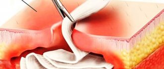
This method is suitable for patients with a disease duration of no more than 1.5-2 years. The essence of this method is to reinnervate the facial nerve (the nerve that conducts the impulse is sutured to the “non-functioning” facial nerve) with another motor nerve (usually the masticatory nerve or hypoglossal nerve is used). The operation is performed using modern microsurgery. This operation is performed in a hospital under anesthesia and is classified as high-tech medical care (HTMC). The fibers of the working nerve grow along the facial nerve, so the impulse reaches the facial muscles and they begin to contract. As a result, facial movements appear and a certain amount of facial expression is restored.
The cross nerve is a nerve autograft taken from the sural nerve (n. suralis) through a mini-incision in the ankle area. This autograft is sutured to the facial nerve on the healthy side and passed through a tunnel (without visible incisions) and sutured into the facial nerve on the damaged side. Through this graft, a nerve impulse passes from the healthy side to the diseased side, causing the muscles on the damaged side of the face to move. In this case, the donor area (leg) does not suffer functionally, the function is not impaired. This method is often used in clinical practice in combination with other treatment methods.
The rehabilitation period takes from 4 months to 2 years. During this period, it is necessary to strictly follow the recommendations: daily myogymnastics, myostimulation, massage.
| Illustration of facial nerve neuroplasty using the masseteric branch of the trigeminal nerve: |
Autologous muscle transplantation (granus muscle, latissimus dorsi)
This method is suitable for patients with a disease duration of more than 1.5-2 years. The essence of this method is to transfer the muscle from the donor area (gracilis muscle - thigh, latissimus muscle - back) to the face, in which the vessels of the muscle flap are sutured into the vessels of the face using a microscope. Thus, this autograft is supplied with blood and “takes root.” The muscle is reinnervated through the masticatory (or hypoglossal) nerve and the cross nerve. This operation is performed in a hospital under general anesthesia, which is also high-tech. The transplanted muscle imitates contractions of the muscles of the middle zone of the face, that is, a smile appears. An invisible thin scar remains in the donor area, the function of which is not impaired.
The rehabilitation period takes from 4 months to 2 years. During this period, it is necessary to strictly follow the recommendations: daily myogymnastics, myostimulation, massage.
| Illustration of lean muscle autotransplantation: |
Autotransplantation of muscles (temporalis muscle).
This method is suitable for patients with a disease duration of more than 1.5-2 years. The essence of this method is to move the temporal muscle without disrupting its blood supply and innervation to the midface area, forming a nasolabial fold. This operation is performed in a hospital under general anesthesia. When this muscle contracts, a smile is formed. Also, this method achieves facial symmetry at rest. The rehabilitation period takes from 4 to 10 months. During this period, it is necessary to strictly follow the recommendations: daily myogymnastics, myostimulation, massage.
| Illustration of temporalis muscle transposition: |
Static facial correction.
This method is suitable for patients with any duration of the disease. Often used as an adjunct to previously described treatment options. The essence of this method is to move and fix the soft tissues of the face on the diseased side into a symmetrical position relative to the healthy side. The result is symmetry of the face at rest, movements do not appear (if this method is used separately, without combination with previously described methods). With this correction, threads, meshes, and fascia are often used for additional tissue fixation. The rehabilitation period takes from 1 to 6 months.
With all methods of surgical treatment, incisions are made, as in a facelift, in the scalp, along the natural fold in the pre-auricular area, behind the auricle. After such incisions there are no visible scars. The donor area, regardless of the method, does not suffer functional losses.
1.What is facial pain and classification of prosopalgia?
Facial pain
(“prosopalgia” from the Greek prosopon - face and algos - pain) - refers to a number of symptoms that cause diagnostic difficulties. The surface of the face and its internal structures have many nerve endings and nodes, and pain in them can be associated with the face itself or other organs and parts of the body.
Since the face contains organs related to several body systems, several medical specialists can study and treat facial pain (ENT, ophthalmologist, neurologist, surgeon, dentist). Facial pain can be part of some syndromes, the symptom complex of which affects not only the facial areas. Facial pain can be unilateral or symmetrical.
According to its genesis, facial pain can be classified into the following groups:
- neuralgic pain (somatalgia);
- pain caused by vascular disorders, for example, migraine (sympathalgia);
- muscle pain;
- soreness of the facial bones;
- skin pain;
- osteochondrotic pain;
- pain of inflammatory origin;
- traumatic and post-traumatic pain.
A must read! Help with treatment and hospitalization!
Results of surgical treatment of paralysis of facial muscles
The result of these operations (with the exception of static correction) does not appear immediately. As a rule, the result appears after 4-8 months - the first weak movements appear, which gradually intensify with mandatory adherence to the specialist’s recommendations. This is due to the fact that nerves re-grow, according to the literature - up to 1 mm per day. First, sensations appear in the form of “lumbago”, “running goosebumps”, etc. This indicates the growth of the nerve along the nerve fibers. The first movements are quite weak, since the muscles have been without load for a long time, so it takes time for their functional restoration, and it is necessary to constantly load them rationally. Thus, rehabilitation plays a very important key role in the final outcome. And the patient must be prepared for daily exercise and not immediate results.
3.Treats for facial pain
When your face hurts, you should not self-medicate until you see a doctor. Even taking painkillers should be postponed, although most often after an examination the doctor prescribes something for the symptomatic treatment of pain. The fact is that the picture and nature of pain are an important diagnostic criterion, so a blurry picture of facial pain will complicate the examination and making a correct diagnosis.
If during the diagnosis it is possible to determine the cause of facial pain, then treatment of the causative disease is prescribed, and, as a rule, the pain goes away when a therapeutic effect is achieved. This does not exclude the use of painkillers at the beginning of treatment, since prosopalgia can be quite painful and lead to disturbances in sleep and performance.
Treatment of atypical pain that does not have a specific cause includes the drugs carbamazepine, finlepsin, mazetol
and others used for trigeminal neuralgia.
In addition, a psychological examination is indicated, identifying the triggers for pain, and psychocorrection sessions. Physiotherapy can also be a good addition
- magnetic and laser therapy;
- electrophoresis with analgin and lidase;
- electrosleep;
- treatment with diadynamic currents;
- hydrocarbon treatment.
About our clinic Chistye Prudy metro station Medintercom page!
Where is surgical treatment for paralysis of the facial muscles performed?
Specialists from the Department of Maxillofacial and Reconstructive Surgery of the Federal State Budgetary Institution NKCO FMBA of Russia, together with the Department of Ear Diseases, have developed and put into practice a unique method of surgical treatment of patients with paralysis of the facial muscles. The first operations on patients with this pathology in Russia were performed by specialists from the Federal State Budgetary Institution NCCO FMBA of Russia - otosurgeon, MD. Hassan Diab and maxillofacial surgeon Ekaterina Orlova.
A healthy person rarely thinks about how important facial expressions are and what role they play in our lives, especially a smile... Smile more often!
Diagnostics
A neurologist is involved in determining the cause of weakness of the facial muscles. During the survey, the specialist finds out the time of symptom onset, identifies other signs, and examines the dynamics of disease progression. To clarify the diagnosis, procedures such as:
- CT scan
. A CT scan of the skull is performed for fractures of the pyramid, jaws, and zygomatic bone. A CT scan of the brain is performed for inflammatory lesions, areas of ischemia or hemorrhage, and structural disorders. - Magnetic resonance imaging
. MRI of the brain is recommended for TBI, strokes and neoplasia. It is performed natively or using contrast, and can be supplemented with angiography. - Electroencephalography.
It is carried out to assess brain functions in tumor processes and post-stroke conditions. - Electrophysiological techniques
. Electromyography, electroneurography and evoked potentials are recommended to determine the level and extent of damage to the facial nerve. During treatment, it is prescribed to assess the effectiveness of therapeutic measures. - Lab tests
. Required to identify pathogens of infectious diseases, determine specific markers of autoimmune pathologies, and establish the nature and degree of differentiation of neoplasia.
Facial taping