Hutchinson, Pfluger and Fournier teeth are a type of dental enamel hypoplasia. This disease appears, as a rule, due to mechanical trauma to the follicles or when infection penetrates into the buds of the teeth. The most common cause is considered to be incomplete development and even absence of tooth tissue. Let's find out how Hutchinson's teeth develop.
Causes of hypoplasia
Often the disease occurs as a result of congenital pathology, although it develops only after the birth of the child. What causes Hutchinson's teeth to develop? The reasons for this are as follows:
- Conflict of Rh factors in the blood of the child and mother.
- Infectious diseases suffered by a woman in the first 3 months of pregnancy.
- Severe and prolonged toxicosis during the 2nd and 3rd trimester.
- Injuries received during childbirth.
- Childbirth that occurred before 40 weeks (premature).
- Rickets.
- Dystrophy of the child (with poor appetite and other reasons).
- Diseases of the gastrointestinal tract.
- Metabolic disorders in the body.
- Somatic diseases.
- Improper brain function in the first year of life.
- Infectious diseases suffered by a child in utero or after birth up to 6 months.
- Injuries to the jaws and face.
Local hypoplasia
- Several teeth are damaged.
- Inflammatory processes may occur due to damage to the deep layers.
- Structural defects appear on the teeth.
- Affected teeth may have some or all of their enamel missing.
In addition to the two main forms of the disease, doctors also distinguish 3 special forms.
These include:
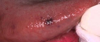
- Hutchinson's teeth. Usually several or all teeth change shape. They take on a round or oval appearance, and their cutting edges become concave and resemble a crescent moon.
- Pflueger's teeth. This form is very similar in appearance to the disease described by Hutchinson. The only difference is the appearance of the cutting edge, which looks the same as that of a healthy person.
- Fournier's teeth. Permanent teeth, namely “sixes,” have the shape of a cone. They are wide from the root and narrow downwards. On their surface there are tubercles that are almost invisible. Often this form develops with syphilis (intrauterine).
Malocclusions
Malocclusions are deviations from the normal relationship of the dentition of the upper and lower jaws. These deviations can be considered in three directions:
Sagittal
Prognathia (distal bite) - characterized by a mismatch in the relationship of the dentition due to the protrusion of the upper teeth or distal displacement of the lower jaw. Distal occlusion may be partial or total; jaw, skeletal or dental; with or without displacement of the lower jaw. Etiology: congenital structural feature of the facial skeleton, childhood diseases affecting the development of the skeletal system, inflammatory processes in the nasopharynx, etc.
Progenia (mesial bite) - characterized by inconsistency of the dentition due to protrusion of the lower teeth or mesial displacement of the lower jaw. It may be partial or complete; jaw, skeletal or dental; with or without displacement of the lower jaw. Etiology: congenital structural feature of the bones of the facial skeleton, incorrect method of artificial feeding, early loss of primary molars, etc.
Transversal
Transversal narrowed dentition - discrepancy between the width of the upper and lower dentition
Vertical
- deep bite - a closure of the dentition in which the front teeth are largely overlapped by the antagonists. Depending on the vestibular or oral inclination, two types of deep bite are distinguished - vertical and horizontal. Etiology: congenital structural feature of the facial skeleton, childhood diseases affecting the growth and development of bones, early loss of primary molars. The main goals of treatment are to separate the bite, expand the narrowed dentition on the jaw that is lagging behind in development, and, if necessary, move the lower jaw.
- open bite - characterized by the presence of a gap between the teeth with central occlusion. This gap occurs more often in the area of the front teeth. There are two forms of open bite - vertical and horizontal. Etiology: rickets, difficulty in nasal breathing, early loss of frontal teeth, wide diastema.
- crossbite - characterized by the reverse closure of the teeth of the right or left half of the bite. Etiology: delay in the replacement of milk teeth by permanent ones, incorrect position of the tooth buds and subsequent incorrect eruption of these teeth, uneven development of the jaws and dental arches.
Degrees of the disease
There are 3 degrees of the disease. They vary in complexity and shape.
- The initial degree of hypoplasia manifests itself in the form of small pigment spots located on the surface of all or several teeth.
- The average degree of hypoplasia manifests itself when convex or concave grooves, as well as pits, appear on the surface of the enamel. Often against this background, Hutchinson's triad develops.
- A severe degree of hypoplasia is observed when tooth deformation occurs or the enamel is erased.
Treatment is available for any degree, but treatment methods vary.
Anomalies of the dentition
Anomalies of the dentition are characterized by a change in the shape of the typical dentition of the upper or lower jaw, which is caused by their narrowing or expansion in various areas and is expressed by crowding of the teeth, their rotation along the axis, vestibular or oral teething, partial edentia, the presence of supernumerary teeth, diastemas and etc. The following irregular forms of dentition are distinguished when they are narrowed:
- acute-angled, when the narrowing is localized in the canine area;
- saddle-shaped, when the narrowing is more pronounced in the premolar area;
- V-shaped, when the dentition is narrowed in the lateral areas, the frontal area acts as an acute angle;
- trapezoidal, when the frontal area is narrowed and flattened;
- common, when all the teeth - frontal and lateral - stand closely together;
- asymmetrical, in which the narrowing is more pronounced on one side of the dentition of the upper or lower jaw, resulting in a crossbite.
The main etiological factors of anomalies in the shape of the dental arches are underdevelopment of the jaws and their deformations caused by diseases of early childhood.
Forms of the disease
Dentists divide enamel hypoplasia into 6 forms:
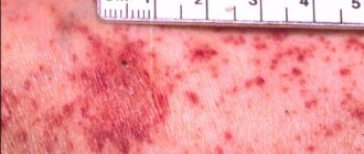
- Spotted. With it, white spots appear on the surface of the teeth, which causes a change in the structure of the tissue. Sometimes the color of the spots may be yellow or light brown. The central incisors become stained first.
- Erosive, or cup-shaped. It appears in the form of round or oval defects, similar to a cup, which differ from each other in size. The erosive form has a paired nature; it often affects teeth located symmetrically. The enamel may thin towards the bottom of the bowl, and sometimes be completely absent. In some cases, the stains may take on a yellow tint due to dentin leaking out.
Problems that pathology leads to
An awl-shaped tooth in a child or adult is not only an aesthetic and psychological problem. A person with this defect can constantly injure the oral mucosa, which in turn is fraught with stomatitis, gingivitis and periodontitis, the formation of painful and poorly healing ulcers and their degeneration into malignant tumors.
An abnormal form can become a prerequisite for the formation of malocclusion, as well as the cause of poor-quality chewing of food, dysfunction of the maxillofacial apparatus, and problems with the gastrointestinal tract.
On a note! If spike-shaped teeth are supernumerary, then they provoke a phenomenon called crowding. When overcrowding occurs, high-quality hygienic oral care is difficult; small pieces of food are poorly removed from the spaces, which leads to increased accumulation of bacterial plaque, the appearance of halitosis, caries and gingivitis.
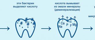
The defect is often accompanied by enamel hypoplasia, which means that hard tissues are very vulnerable and sensitive to any external influences and bacteria. The enamel in patients with this problem is quickly destroyed and is susceptible to caries and its complications (pulpitis, periodontitis).
Diagnosis of dental hypoplasia
It is quite easy to identify the disease, especially in the later stages. However, in the early stages the disease can be confused with initial and superficial types of caries.
| Symptom | Caries | Hypoplasia |
| Stains | A single white spot is located on the surface near the neck of the tooth. | Multiple spots are white or yellow-brown in color and are located over the entire surface of the tooth. |
| Enamel condition | The enamel has a smooth and even surface. | The enamel surface is covered with grooves and pits, and in rare cases may be partially or completely absent. |
| Form | The teeth have an unchanged shape. | In some types of disease, the teeth are modified, have a barrel shape, and the cutting edge resembles a crescent. |
If you find signs of illness, consult a doctor and he will make an accurate diagnosis.
Treatment
If the hypoplasia is weak and there are stains on the teeth that are invisible to the naked eye, then treatment may not be necessary. When stains are noticeable or the process of tooth decay has begun, it is necessary to urgently consult a doctor, who will immediately take appropriate measures. No matter how sad it may sound, the disease cannot be cured completely. Dentists can correct cosmetic defects, but there is a chance that after a while you will have to contact them again.
The main treatment is teeth whitening. This helps remove stains from the enamel. However, this method is not used in severe stages of the disease. Sometimes doctors grind teeth, which helps get rid of bumps and uneven cutting edges.
Preparing teeth for prosthetics using the INSIGNIA system
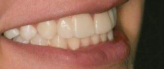
Problem: a young woman came to the Dial-Dent family dental clinic with a desire to correct the incorrect position of her front teeth and improve their overall appearance. She did not like the gaps between the teeth and the tenon-shaped upper incisor (second from the right), which was not only unsightly, but also incorrectly positioned.
Decision: orthodontist E.V. Rada prepared for prosthetics by correcting the bite using the INSIGNIA brace system. Orthodontic treatment pursued the following goals: closing the gap between the teeth , eliminating crowding of the teeth below, and also freeing up space for prosthetics of the second upper incisors. Orthopedic doctor A.S. Ivankov performed prosthetics of the second incisors using crowns.
The initial consultation includes an examination, analysis of the current clinical picture and general recommendations from the orthodontist. To draw up a complete treatment plan, more thorough research and calculations of the data obtained are necessary: the patient needs to take a panoramic photograph of the teeth, TRG in the lateral projection, and also visit an osteopath, speech therapist, or dental therapist (all this can be done at the Dial-Dent clinic). The orthodontist will take impressions of the patient’s teeth and photograph the patient from different angles for further evaluation of the treatment. Additional consultations with the above-mentioned specialists are associated with Dial-Dent’s comprehensive approach to the treatment of orthodontic patients. An osteopath relieves pressure, tension and spasms in the head and neck area that are associated with orthodontic problems. A consultation with a psychologist is necessary to determine the patient’s motivation and check his readiness for long-term orthodontic treatment. You cannot begin treatment if the patient is not sure of the result, is not ready to wear braces for a long time, is too tense and is under stress. Treatment by a dentist is necessary to eliminate dental caries before installing braces, because while wearing braces, hygiene can deteriorate and untreated caries can spread to other teeth. In addition, at the Dial-Dent clinic, all patients in the orthodontic department undergo professional teeth cleaning before installing braces. The orthodontist brings all the data together and draws up a treatment plan.
Photo before orthodontic treatment:
The diagnosis was made by orthodontist E.V. Rada: neutral bite, first skeletal class, protrusion of the incisors of the upper jaw, diastema on the upper jaw, short wide frenulum of the upper lip , anteinclination of the jaws, close position of the incisors of the lower jaw, anomaly of the shape of the teeth: 12 - spinous and 22 - microdentia. To fulfill all the wishes of the patient, the comprehensive work of the following doctors is needed: an orthodontist (normalization of the bite, alignment of teeth, preparation of a place for dental prosthetics), a surgeon (plastic of the frenulum of the upper lip), an osteopath (relieving muscle tension, stabilizing treatment results) and an aesthetic dentist (correction shapes of incisors 12, 22 using crowns).
The orthodontist suggested that the patient remove the tenon tooth (12) and restore it with an implant. However, the patient insisted on temporarily preserving the tooth, and she planned implantation at a later date, so the shape of the incisors will be corrected using crowns on the existing teeth.
Orthodontic treatment was performed using the Insignia brace system . A distinctive feature of Insignia from traditional braces systems is the individualization of braces and computer modeling of the process and result of treatment. The position of the braces is calculated according to the treatment plan. Installation of braces is carried out using a special mouth guard, which allows you to perform this procedure as accurately as possible. Insignia is a progressive system that allows you to get excellent results in a shorter period of time compared to traditional braces systems.
Modeling the result, the orthodontist left equal spaces between the central incisors and canines, so that in the future, when the patient wants to remove the tenon tooth and install an implant, full-fledged (more precisely, full-size, and therefore more aesthetic) prosthetics could be performed. A tenon tooth is too narrow, and the crown on it will differ slightly in size from the crown on a symmetrical incisor. The setup model of the result was discussed with the patient. Insignia braces and arches are made from it within 1-2 months .
Braces are installed on the teeth. The patient visited the orthodontist every two months. The surgeon performed frenuloplasty some time after the installation of braces, when the diastema was closing. The duration of wearing braces was 16 months.
The treatment process is not completed at this point, the patient continues to wear a dental guard (upper jaw), and the doctor installed a permanent retainer on the lower jaw. The retention period is an important component of orthodontic treatment, stabilizing its results.
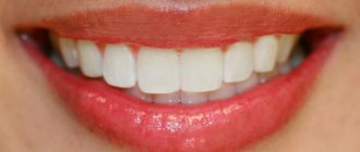
After the braces were removed, aesthetic dentist A.S. joined the treatment. Ivankov, who performed prosthetics of the second incisors with crowns. As noted above, a tenon-shaped tooth has a small neck diameter and with high-quality prosthetics, the doctor cannot significantly expand the crown so as not to lose the reliability of the structure. For this reason, there is a small gap between the teeth, which will be completely closed with full prosthetics using an implant.
The results of the treatment fully correspond to the set goals: correction of the bite and alignment of the teeth was completed successfully, the gap between the teeth was eliminated, the shape of the teeth was corrected, the front teeth have a harmonious appearance.
The cost of orthodontic treatment ( Insignia ) was 112,200 rubles (find out current prices at a face-to-face consultation).
See other examples of orthodontic treatment from Dial-Dent specialists here.
Nutrition
Proper and balanced nutrition plays an important role in prevention. It must be observed even at the stage of pregnancy planning. Also, the child’s nutrition needs to be monitored after birth. When doctors allow your baby to eat new foods rather than milk and formula, the main thing is to include the following in his diet:
- Milk, cheese, cottage cheese and other products that contain calcium and fluoride.
- Vitamin D. You can give your child special medications and spend more time in the sun.
- Foods rich in vitamin C. These are broccoli, oranges, tangerines, spinach.
- Products containing vitamins A and B. These include seafood, legumes, poultry and mushrooms.
Tips for parents
Many parents do not even suspect that dental hypoplasia is a very common disease among children. In order to alleviate the child’s condition, the following actions must be taken:
- Eliminate all sour and sweet foods from your diet.
- Use special toothpastes.
- For small children, purchase silicone finger brushes for oral hygiene.
- Carry out the procedure of silvering your teeth regularly.
- Monitor their condition and promptly fill teeth if necessary.