The zygomatic bone is one of the paired elements of the skull structure, consisting of dense spongy-type plates. The fragility of these areas causes a high probability of fracture under active mechanical stress, the consequences of which depend on the complexity of the deformation. Simple cases are characterized by the absence of displacement of the fragments, as well as the preservation of the bone structure against the background of damage to the arch itself. However, any damage of this kind requires medical intervention, excluding negative consequences for the health of patients.
Symptoms of manifestation
Bruises on the face are difficult to ignore. It is characterized by knowledge of all standard symptoms, such as:
- Painful sensations in the facial area. And they are usually felt strongly because the ends of the facial nerves are among the most sensitive;
- Tissue swelling. This manifests itself as swelling of the skin, which may appear thickened upon palpation. The severity of edema depends not only on the severity of the injury, but also on the thickness of the skin and the internal structure of the tissues. Therefore, the area around the mouth and face is most susceptible to swelling;
- Bruises, bruises and hematomas. They arise as a result of damage to blood vessels and the accumulation of platelets in the damaged area. It’s worth saying right away that the deeper they are under the skin, the later the reaction will appear, but, unfortunately, the longer it will take. For this reason, many people start using ointments and gels for bruises even before visible signs appear;
- Feeling numb. This occurs in cases where the fibers of the facial nerve were directly affected during the injury. If nerve damage is very severe, there is a risk that reduced nerve activity may persist;
- Disruption of various areas of the face. Examples include: inability to see if the eye is swollen, difficulty breathing if the nose is injured, difficulty chewing food if the jaw is injured;
- Open bleeding. Observed when the skin is damaged at the site of the bruise and there is an open wound or deep scratch;
- Nausea, vomiting, loss of consciousness, convulsions. Such serious symptoms can occur if a bruise of the soft tissues of the face is accompanied by a traumatic brain injury and, as a consequence, impaired brain function.
Bruises and bruises are symptoms of contusion of the soft tissues of the face.
Of course, each of these symptoms may vary in severity in each individual case. Much depends on the individual characteristics of the body, such as the thickness of the skin or the elasticity of blood vessels. So, with one type of injury, one person may only have swelling, while another may have a severe hematoma.
Isolated zygomatic arch fractures: our treatment experience
Key words: zygomatic arch, lateral midface, trauma, zygomatic complex
Isolated fractures of the zygomatic arch are a fairly common pathology. In maxillofacial traumatology, their share ranges from 6.5 to 20%, according to various domestic and foreign authors [1,5]. The treatment of these fractures has received and still receives close attention due to the complex anatomy of this area and the frequent occurrence of this pathology.
The purpose of this study is to present our method of treating isolated zygomatic arch fractures, based on many years of experience in treating patients in this category.
Isolated fractures of the zygomatic arch occupy the largest volume among similar injuries of the lateral midface. According to domestic and foreign authors, they account for 12.2–14.8% [1,3], among our patients they accounted for 15.5% of all fractures of the lateral midface.
This feature is associated with the location of the zygomatic arch and its significant fragility. These fractures mainly develop as a result of lateral impacts to the arch area at a right angle. As a rule, they are the result of domestic injuries and road traffic accidents.
Isolated fractures of the zygomatic arch occur with the fragment displaced inward; less often, the fragment is displaced downward. Patients' complaints mainly boil down to tissue retraction in this area - the development of deformation of the zygomatic region, as well as difficulty or impossibility of opening the mouth.
In case of fresh fractures in the absence of post-traumatic edema, the sinking of the arc section and sharp bone protrusions are determined by palpation.
When the zygomatic arch is fractured and its posterior parts fall inward, the mobility of the coronoid process of the mandible and the temporal muscle is impaired, which makes the full range of movements in the joint impossible. In some cases, lateral movements are difficult or there is no complete closure of the teeth. This clinical picture is typical in 87–88% of cases of isolated fractures of the zygomatic bone [4].
Diagnosis of these injuries is based on a carefully collected history, clinical examination of the patient and auxiliary x-ray diagnostics.
The most used x-ray examination in such cases is a simple x-ray of the skull in a lateral semi-axial projection. A radiograph in semi-axial projection is also informative.
There are a wide variety of methods for reducing the zygomatic arch. They come down to conservative and surgical.
One of the very first methods for repositioning the zygomatic bone and arch with fresh fractures is the Gillis technique, proposed back in 1927. and further improved by Kilner and Stone. The essence of the technique is that access is created to the zygomatic bone and arch from the temporal region through an incision of the skin and subcutaneous fat about 2 cm in length, slightly receding inward from the border of the hairline. A long, wide elevator is inserted into the incision, which moves under the zygomatic bone and arch, and thus their reposition is carried out.
However, this technique is quite traumatic, and in most cases, preference is given to intraoral methods of reducing fragments.
Domestic specialists most often use the Limberg-Bragin method. Single-prong hook A.A. Limberg or two-pronged hook Yu.E. Bragin is introduced through a puncture incision 0.3–0.5 cm long in the area of the projection of the lower edge of the zygomatic arch. Using an outward movement, the fragments are set, bringing the hook under their ends shifted inward.
With this technique, the question of fixation of fragments remains open, which in some cases (12.3–13.0%) leads to postoperative displacement of fragments if patients do not comply with the regimen [6].
After careful processing of domestic and foreign literature, as well as based on our own many years of clinical experience, we use the technique we have developed to treat patients with fresh fractures of the zygomatic arch.
A thick curved needle with a strong polyamide thread is passed under each fragment of the zygomatic arch and, by pressing on the thread, the fragments are set and fixed using the same thread to a plate made of brass or aluminum, previously made and modeled in size corresponding to the length of the arch. To prevent bedsores, iodoform turunda is placed under the plate in 2–3 layers. The fixation is removed after 8–10 days.
The advantage of this technique is the low traumatic nature of the manipulation and the possibility of fixing fragments using a plate.
Clinical example.
The victim V., 45 years old, was hospitalized in the clinic of maxillofacial surgery of the 8th med. association of Yerevan 02/05/2005 with complaints of pain in the zygomatic region on the right, impaired facial configuration, difficulty opening the mouth.
According to the patient, the injury occurred as a result of a traffic accident. He did not lose consciousness, did not notice nausea or vomiting.
Clinical and radiological examinations established a diagnosis of an isolated fracture of the right zygomatic arch.
On 02/05/2005, under anesthesia, a surgical intervention was performed - repositioning the zygomatic arch using the described method.
The postoperative period proceeded smoothly. Functional and anatomical rehabilitation developed within 4–5 days. The duration of inpatient treatment is 4 days.
Rice. 1. Zygomatic arch before reposition
Rice. 3. Fixation of the zygomatic arch
Rice. 2. Zygomatic arch after reduction
Conclusions:
- This technique is less traumatic,
- This technique allows not only repositioning the zygomatic arch, but also its reliable fixation,
- The technique allows to reduce the rehabilitation time of patients.
Literature
- Aleksandrov N.M., Arzhantsev P.Z., Vikhriev B.s. and others. Injuries of the maxillofacial region. M.: Melitsina, 1986, ts. 448.
- Gosain AK, Song L., Corrao MA, Pintar FA Biomechanical evaluation of titanium, biodegradable plate and screw, and cyanoacrylate glue fixation systems in craniofacial surgery, Plast. Reconstr. Surg., 1998 Mar; 101(3): 582-91
- Lee CH, Lee C, Trabulsy PP et al. A cadaveric and clinical evaluation of endoscopically assisted zygomatic fracture repair, Plast. Reconstr. Surg., 1998 Feb; 101(2): 333-45; discussion, 346-7.
- Orringer JS, Barcelona V., Buchman SR Reasons for removal of rigid internal fixation devices in craniofacial surgery, J. Cranofac. Surg., 1998 Jan; 9(1): 40-4
- Patel BC, Patipa M, Anderson RL et al. Management of postlepharoplasty lower eyelid retraction with hard palate grafts and lateral tarsal strip, Plast. Reconstr. Surg., 1997 Apr., 99(5): 125-60
- Rohrich RJ, Watumull D. Comparsion of rigid plate versus wire fixation in the management of zygoma fractures; a long-term follow-up clinical study, Plast. Reconstr. Surg., 1995 Sep; 96(3): 570-5
- Zachariades N., Papademetriou I., Rallis G. Complications associated with rigid internal fixation of facial bone fractures. J. Oral. Maxillofac. Surg., 1993 Mar; 51(3): 275-8; discussion, 278-9.
- Zingg M., Laedrach K., Chen J. et al. Classification and treatment of zygomatic fractures: a review of 1,025 cases, J. Oral. Maxillofac. Surg., 1992 Aug; 50(8): 778-90
First aid
First aid is recommended for injuries to the soft tissues of the face, regardless of the force of the blow. Here are three basic steps in this situation:
- Place something cold on the bruised area. This could be ice, snow, a compress soaked in cold water, or even a chilled metal spoon. Cold helps constrict blood vessels, reducing the risk of bruising under the skin. However, there are two things to keep in mind. Firstly, facial skin is quite thin and cannot be exposed to prolonged exposure. The optimal time is 15-20 minutes. The procedure can be repeated after 2 hours. Use material that protects the receptors from hypothermia. The second point is that the effectiveness of this method is available only in the first half hour after injury;
- If there are signs of skin trauma, be it wounds or abrasions, it is important to treat them with antiseptics. The most common of them are the well-known hydrogen peroxide, herbs, and a very weak solution of manganese solution. This must be done very carefully, since every touch to the wound can cause pain to the victim;
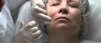
- Anesthesia. If the sensations are strong, you should turn to painkillers. Among them are Ketone, Ketorol, Ibuprofen. Before use, be sure to read the instructions.
First aid for bruised soft tissues of the face includes applying ice.
In most cases, the listed first aid measures for bruises are sufficient. However, if the injury is much more serious and the victim experiences heavy bleeding or convulsions, then the first step is to call an ambulance. When traveling, control bleeding with a tourniquet or position the person so that they do not risk swallowing their tongue.
It is important to remember that the method of providing first aid will affect the result of further treatment of facial bruises and the likelihood of developing complications.
First aid
In a situation with a serious injury, the people around him must provide first aid to the victim. Medical recommendations exclude independent adjustment of individual parts of the skull or contact with open wounds. The main tasks are to create conditions of complete rest, eliminate re-injury, try to stop the bleeding and call an ambulance team.
In case of acute pain, an injection of an anesthetic is allowed. To stop continuous bleeding, it is necessary to press down the artery. In cases where the wound is small, you can treat the area with hydrogen peroxide, as well as ensure careful application of cold.
Diagnostics
A bruise is easy to diagnose, but only doctors can determine its severity and possible complications. Therefore, if for any reason the injured area is bothering you, it is better not to waste time and seek professional help. The diagnosis can be made:
- inspection;
- palpation;
- Assessment of symptoms and complaints;
- In some cases, an ultrasound or x-ray examination may be required.
Diagnosis consists of determining not only the presence of a bruise, but also its severity. There are four degrees:
- First degree. This is the easiest and least dangerous option, involving a slight change in the structure of the subcutaneous tissue. There is no bleeding or hematoma, but blue skin discoloration is quite possible. For the first degree of injury, visiting a doctor is usually not required. It is quite possible to limit yourself to home remedies, which, with the right approach, will help eliminate symptoms in 5-7 days;
- Second step. In this case, significant damage to the subcutaneous and muscle tissue occurs. Severe swelling, pain and even hematoma may appear. It is better to treat such a bruise with medications together with physiotherapeutic procedures;
- Third degree. It damages not only muscle tissue, but also tendons. In some cases, the integrity of the skin may be compromised. Since there is a risk of developing an infectious process, examination by a doctor is mandatory.
- Fourth degree. This is the most severe and dangerous degree of injury. It is always accompanied by injuries not only to soft tissue, but also to bone tissue. In this case, there is a high risk of developing various types of complications. It is imperative to seek qualified medical help.
Accordingly, the type of treatment will directly depend on the degree and nature of the bruise.
Bruises and hematomas of the face.
Bruises are the result of a mild blow to the face with a blunt object. In this case, subcutaneous fatty tissue (SFA) and facial muscles are damaged without tearing the skin. Hemorrhage occurs and pronounced post-traumatic tissue swelling appears, that is, a superficial or deep hematoma is formed.
Deep hematoma - blood enters the interstitial space forming a cavity.
Superficial hematoma - imbibition (soaking) of tissues with blood occurs without the formation of a cavity. The nature, color and time of resorption of the hematoma depend on its location, depth and size of the damage. The outcome of hematomas is most often favorable; they completely resolve without leaving any trace.
The nature, color and time of resorption of the hematoma depend on its location, depth and size of the damage. The outcome of hematomas is most often favorable; they completely resolve without leaving any trace.
Bruises, hematomas and abrasions are the mildest types of injury, but are they so harmless?
Treatment
Treatment methods for bruises can be divided into two large groups: traditional in the form of drugs and treatment methods, and non-traditional in the form of traditional medicine. Ideally, you can combine techniques from both groups. This solution will help you get rid of bruises in less time.
Medication
If the situation does not require hospitalization, the bruise is treated using standard methods. Namely:
- Preparations for external use: gels, ointments, creams;
- Physiotherapeutic procedures. These include electrophoresis, heating, laser therapy. All these procedures are aimed at eliminating subcutaneous clots and stimulating skin regeneration.
Creams, ointments, and other treatments for bruises are widely available.
In every pharmacy you can find a huge selection of such products. But when choosing, it is important to pay attention to the composition, influence and age restrictions. Not all products are suitable for children.
With regular and correct use of the ointment, you can get rid of bruises and swelling within a few days. And if you supplement your treatment with physical therapy, you can expect results even faster.
Traditional methods
Bruises and bruises can also be treated with traditional medicine. The only rule is that you can start using them a few days after the injury.
Among the folk recipes for bruises and bruises on the face it is worth mentioning:
- Cabbage leaf or raw potato. They should be applied to the damaged area. Thanks to this, swelling quickly decreases;
- Honey. Has a good laxative and anti-inflammatory effect. You can even not limit yourself to the area of the bruise, but apply honey all over your face, creating the appearance of a mask;
- Camphor oil. To obtain the effect, it must be applied with light rubbing movements;
- Compresses. It is best to cook them with onions or salt;
- Alcohol-based compresses. In this case, the rosemary plant can be the basis. Has a warming and antiseptic effect;
- Arnica decoction. This measure is applied not externally, but internally. It not only strengthens the immune system, but also stimulates regenerative processes.
Applications with honey are a folk method for treating bruises of the soft tissues of the face.
And, of course, one should not underestimate the influence of standard massage measures in the form of light stroking and rubbing.
Consequences
Complications with bruises are quite possible. It all depends on the nature of the injury and which area of the face was damaged. Possible consequences include:
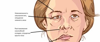
- Nerve damage. completion. This is quite dangerous, since it is not always possible to fully restore their functionality. This means that the affected part of the face may stop moving;
- Deterioration of vision. If the area around the eyes is bruised, there is also the possibility of damage to the nerve responsible for visual functions. Again, the outcome depends on the nature of the injuries. Both partial and complete loss of vision is possible, which is much less common;
- suppuration at the site of the bruise in the form of an abscess;
- bleeding that, if not treated immediately, may lead to fainting or shock;
- Cyst formation in the presence of hematomas.
In case of serious injuries, the bruise may be accompanied by a concussion, deformation of the bones of the nose or jaw. As a result, a person may later suffer from conditions such as sinusitis or sinusitis.
Prevention
Almost all of us have had bruises on the soft tissues of our faces in our lives. Unfortunately, this cannot be completely prevented. However, in order to at least minimize the risk, it is necessary to take basic precautions and safety measures. When it comes to children, it is important to teach them from an early age how to avoid traumatic situations.
If facial bruises do occur, do not leave it unattended and take appropriate measures. You can do this yourself or seek medical help.