131
The impeccable functioning of the human dentofacial apparatus is possible only with the complete integrity of the jaw rows.
The absence of even one element triggers a process of negative morphological and functional changes, which become increasingly worse over time.
These changes can be prevented only through timely prosthetics of the defect. Delaying it can lead to serious consequences, information about which is brought to your attention in the article below.
General information
Dental pathology is not a serious disorder, but in the absence of proper treatment it can provoke some complications. The main reason for its development is considered to be the loss of teeth without subsequent implantation or prosthetics. According to research, more than 30% of patients who have had at least one tooth removed do not think about the implantation procedure.
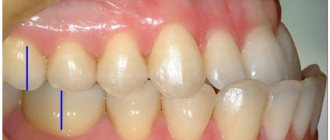
This is how the phenomenon manifests itself when a tooth is lost
This is due to the fact that with such a minimal loss, the process of chewing food is not disrupted, so a person does not rush to the dentist. It is noted that 2-3 months after the removal of one or more units, a gradual displacement of healthy teeth begins. If a person does not visit a specialist for a year to install implants, the procedure will be quite problematic.
When one tooth is removed, the pathology develops quite slowly. If the number increases, rapid displacement of healthy teeth begins, elongation of the alveolar processes and other disorders:
- dysfunction of the muscles of the lower jaw;
- advancement of the upper or lower dentition;
- changes in the structure of the alveolar processes;
- exposure of the root of healthy units located directly next to the void;
- formation of a gum pocket around healthy teeth;
- dysfunction of the temporomandibular apparatus;
- narrowing of the periodontal fissure;
- changes in collagen fibers;
- dystrophic processes in the pulp;
- disturbance in the structure of the periodontium.
As a rule, several months or even years after the removal of one or several units, prosthetics and implantation become very complicated. It is for this reason that experts strongly recommend making a replacement no later than 10-14 days after extraction.
Abstracts on orthostomy (for practicing lectures) / Deformations
Ministry of Health of the Russian Federation
St. Petersburg State University
them. acad. I.P. Pavlova.
Department of Orthopedic Dentistry and Materials Science with a course in Adult Orthodontics
Dental deformation resulting from partial loss of teeth. Popov-Godon phenomenon. Clinic, diagnosis, treatment.
Ivanopulo Modest Ruvimovich
The appearance of defects in the dentition leads not only to a disruption of the morphological unity of the dentition, but also to a complex restructuring that occurs first near the defect and then spreads to the entire dentition. Externally, this restructuring is manifested by the movement of teeth, which often leads to disruption of the occlusal surface of the dentition, i.e. to secondary malocclusion deformities, complicating the clinical picture of partial tooth loss, making it difficult to choose and carry out orthopedic treatment.
The dental arch, as part of the dentofacial system, is a single whole due to the presence of interdental contacts and the alveolar process, in which the roots of the teeth are fixed. The loss of one or more teeth disrupts this unity and creates new conditions for the functional activity of the masticatory apparatus.
The most common causes of tooth loss are caries and periodontitis. trauma, surgery, vitamin deficiency, etc. The resulting clinical picture depends on the number of lost teeth, the location and extent of the defect, the type of bite, the state of the supporting apparatus of the remaining teeth, the time that has passed since the loss of teeth, and the general condition of the patient.
The clinical picture depends on the type of movement. There are two clinical forms of vertical movement of teeth with the loss of antagonists (L.V. Ilyina-Markosyan, V.A. Ponomareva). At the first form
tooth movement is accompanied by an increase in the alveolar process (dental-alveolar lengthening, without a visible change in the height of the clinical crown of the tooth).
This form is typical for tooth loss at a young age. In the second clinical form,
tooth loss occurs with exposure of part of the root, which indicates a later stage of restructuring.
In the second clinical form, two subgroups are distinguished:
subgroup
- visible increase in the alveolar process with slight (within line A) periodontal resorption;
subgroup
- there is no increase in the alveolar process; resorption of periodontal tissue is detected at a level of half or more.
Popov-Godon phenomenon -
displacement of teeth in different directions after the formation of a defect in the dental arch, leading to deformations of the occlusal curve. A complication that develops after the removal of part of the teeth can occur at any age.
With a defect caused by the loss of the main and lateral antagonists, a change in the position of the teeth in the vertical direction is most often observed. A tooth devoid of antagonists appears to be part of a dentition defect; the distance between its occlusal surface and the alveolar process of the toothless area of the opposite jaw decreases, or the teeth touch the mucous membrane.
Studies of the first form of deformity (without exposure of the root) have shown that, despite the increase in the alveolar process, there is no visible addition of bone substance, but a rearrangement of the bone trabeculae occurs.
Based on morphological data, it was concluded that the secondary deformations observed in the clinic are based on the process of restructuring of the tissues of the tooth and jaw due to the loss of their usual functional load! This is an expression of the adaptation of the dentofacial system to new functional conditions.
Partial secondary tooth loss, complicated by the Popov-Godon phenomenon, should be differentiated:
from partial loss of teeth, complicated by a decrease in occlusal height and distal displacement of the lower jaw;
from partial loss, complicated by pathological abrasion of hard dental tissues (localized form, decrease in occlusal height);
3) from partial loss of teeth on both jaws, when not a single pair of opposing teeth has been preserved.
To distinguish the Popov-Godon phenomenon from these forms of partial loss and complications, it is necessary to examine the relationship of the dentition in the position of the lower jaw in a state of physiological rest. To do this, after determining the central relationship of the jaws, diagnostic models are fixed in the articulator and the severity of the occlusal curve is examined both in the anterior section and in the area of chewing teeth, the amount of space between teeth devoid of antagonists and the alveolar process of the edentulous area.
For the purpose of differential diagnosis and treatment, it is recommended to use therapeutic and diagnostic mouth guards aimed at restoring the occlusal height and normalizing the ratio of the elements of the temporomandibular joint.
The study of diagnostic jaw models is one of the main examination methods, the purpose of which is to identify the nature of the occlusal relationships.
Diagnostic models should be made before, during and after treatment.
When assessing diagnostic models, it is necessary to clarify the type of bite, the depth of the incisal overlap, the nature of the closure of the palatal and lingual cusps, etc., as well as the magnitudes of dentoalveolar elongation, the nature of the occlusal curve, the relationship of individual teeth to the mucous membrane of the edentulous alveolar process, the nature of the medial or distal movement teeth, supercontacts, where blockage of lower jaw movements occurs, level of tooth shortening, etc.
The false Popov-Godon phenomenon is mistaken for the true one, since when the jaws are arbitrarily closed without taking into account the height of the lower part of the face, a false impression is created that teeth devoid of antagonists have shifted into the dentition defect of the opposite jaw.
In the process of making a differential diagnosis, one should remember about the possible combination of the Popov-Godon phenomenon with other diseases of the dentofacial system. So. As a result of the loss of all chewing teeth in the lower jaw, the following complications can simultaneously develop: deformation of the occlusal curve, decreased occlusal height and distal displacement of the lower jaw.
In this case, after restoring the height of the lower part of the face and the central relationship of the jaws using wax bases with occlusal ridges both in the mouth and on diagnostic models, the degree of tooth displacement towards the defect is significantly reduced.
Rational prosthetics is impossible without eliminating occlusal disorders, which, in turn, can cause functional impairment.
tions of the temporomandibular joint, functional overload of the teeth, blockade of movements of the lower jaw, etc.
Elimination of occlusal disorders has preventive and therapeutic purposes.
Therapeutic goals are:
normalization of occlusal deviations;
eliminating blocking movements of the lower jaw;
eliminating functional overload of periodontal teeth;
normalization of the function of the temporomandibular joint;
creating conditions for the manufacture of a rational prosthesis design.
Prevention consists of preventing:
functional overload of periodontal teeth;
dysfunction of the TMJ;
dysfunction of the masticatory muscles.
Normalization of the occlusal relationships of the dentition
is achieved:
by grinding off the cusps of the displaced teeth;
shortening of teeth that interfere with the reconstruction of the occlusal plane, if necessary, with their depulpation;
restoration of the height of the lower part of the face;
application of special prostheses that cause restructuring of hypertrophied areas of the alveolar process (hardware or orthodontic method);
application of special prostheses causing restructuring of the alveolar process, with preliminary compactosteotomy (corticotomy) (hardware-surgical method);
removal of teeth, if necessary with resection (alveolotomy) of part of the alveolar process (surgical method);
The choice of method is determined by the nature of the clinical picture, the shape and degree of deformation, age and general condition of the body.
Method
for grinding hard tissues.
This method is used in the treatment of people over 35-40 years old when the teeth are displaced beyond the occlusal plane by no more than half the vertical size of the tooth (teeth). Indications for grinding are the second form of the Popo-va-Godon phenomenon and the unsuccessful use of the disocclusion method.
In order to determine the degree of grinding, diagnostic models or lateral extraoral X-rays are studied, the extent to which the tooth has shifted is determined, which determines the amount of tissue removed from the occlusal surface. If necessary, dental pulping is performed.
After grinding non-pulp teeth, it is necessary to conduct a course of fluoride prophylaxis. If during grinding it is necessary to remove part of the dentin, then it is recommended to make a crown at the same time.
Symptoms and classification
Depending on the nature of the displacement with the Popov-Godon symptom, experts identify several options for the development of the pathology: vertical protrusion of the lower or upper jaw, displacement of the lower or upper teeth, inclination of the teeth or the entire row inward, or a combined disorder. The last type is the displacement of teeth in different directions and to varying degrees.
There is also another classification from V. A Ponomareva. It involves dividing pathology into two forms:
- The first is characterized by tooth displacement and simultaneous lengthening of the alveolar process. The difference is the absence of a gum pocket. The root is also not exposed, there are no dystrophic changes in the pulp. This form is considered mild and it is possible to get rid of clinical manifestations quite quickly.
- The second is accompanied not only by displacement of teeth, but also by the development of an inflammatory process, exposure of the root, and signs of dystrophic changes in the pulp. It is worth noting that with this type, the alveolar process may lengthen or not, depending on the characteristics of the individual patient and the time over which the pathology develops. Treatment in this case is complicated and requires an integrated approach aimed at eliminating the inflammatory process and correcting the dentition.
As a rule, the pathology is accompanied by malocclusion, a decrease or increase in the distance between the teeth, and their deviation forward or backward. There are signs of gum inflammation, tooth root exposure and changes in the pulp. The last symptom appears later than the others and is considered the main one in Popov-Godon syndrome.
General concept
The Popov-Godon phenomenon (P-G) is a pathological restructuring of the dentoalveolar processes due to tooth loss or, in other words, a violation of the integrity of the rows.
The pathology got its name from the dentists who first described it. Namely, the Russian scientist O.V. Popov, who observed in 1880 the deformation of the dentition of a guinea pig, whose incisors were removed, and Godon, who in 1904 described the deformations of the dentofacial apparatus of patients after the loss of elements of the jaw rows.
In addition, Godon is also known for proposing his hypothesis to explain the development of the phenomenon, which he called the theory of articular equilibrium.
The clinical picture of the P-G phenomenon is varied. One of the most easily recognizable manifestations of pathology is the vertical extension of the unit located opposite the defect from the alveolar process, its crown crossing the occlusal plane and entering the space of the defect.
In some cases, advancement continues until full contact with the surface of the gum of the opposite row. Units that are located next to the defect can tilt towards it, rotate around their axis, and shift in one direction or another.
The relationship between different types of pathological displacements in the Popov-Godon phenomenon made it possible to establish a study conducted on 120 young patients who had anomalies caused by adentia.
It turned out that medial displacement accounts for 45.1%, combined - 17.5%, palato-oral - 10%, distal - 9.5%, vertical - 9.5% described above, vestibular - 5 .5%, for tortoposition (rotation around an axis) – 2.2%.
In addition to visual, clearly visible manifestations of pathology, there are also hidden or less visible ones. They are expressed by a violation of the occlusal relationships of individual units, deformation of the occlusal plane, dentoalveolar enlargement and some other features.
The specific form of manifestation of the P-G phenomenon depends on a number of factors:
- Length and location of the defect.
- Time elapsed since tooth loss.
- Age of the patient (negative manifestations occur faster in children).
- General health.
- Conditions and characteristics of the jaw apparatus.
Delay in treating the P-G phenomenon threatens the patient with many negative consequences:
- Chewing dysfunction.
- Deterioration of smile aesthetics.
- Chronic articulation injury, fraught with the risk of malignancy.
- Blocking low frequencies.
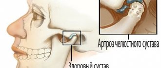
- TMJ dysfunction due to overload.
- Impaired articulation and diction.
- Pain and discomfort in some clinical situations.
- Deterioration of treatment and prosthetic conditions.
The root cause of the Popov-Godon phenomenon is tooth loss, which can occur for various reasons - due to caries, periodontitis and other diseases, injuries, forced extractions.
As a secondary cause of displacement during edentia, one should consider the restructuring of the dentoalveolar process according to mechanisms that are still not completely understood.
There are different theories explaining this process. In particular, Hodon’s theory, mentioned above, is that the jaw apparatus is a system that maintains immobility with the integrity of the rows due to a closed chain of forces.
If at least one tooth is lost, the closed chain breaks, which causes multidirectional displacement.
There are other theories that explain the P-G phenomenon, but there is still no one that would be recognized by all experts.
Let's talk about the impact of bite on the face and the possibilities to correct the situation.
Come here if you are interested in the connection between bite and headaches.
At this address https://orto-info.ru/zubocheliustnye-anomalii/okklyuzii/ispravleniya-prikusa-viniryi-v-stomatologii.html find out whether veneers correct the bite.
Diagnostic methods
To determine the pathology, a specialist examines the oral cavity and assesses the condition of the lower and upper jaw. To accurately determine the shape, it uses the classification of the Popov-Godon phenomenon. It is with its help that the doctor will be able to more accurately determine the stage of development of the disease.
An important point is differential diagnosis, which helps to separate pathology from partial edentia, which has similar clinical manifestations. For this purpose, the specialist compares the ratio of the dentition to each other in a state of physiological rest. It is also necessary to determine the distance between the teeth in the anterior and lateral projections.
The first parameter is considered the most important, since only when a displacement is detected in the central region can we talk about the development of a pathological process. If clinical manifestations are present, but there are no changes in this area, edentia of varying degrees of severity is most often suspected.
An additional method is an X-ray examination and study of the resulting images. After establishing an accurate diagnosis and the extent of the lesion, the specialist determines a treatment regimen that is individual in each case.
What is the Popov-Godon phenomenon?
The Popov-Godon phenomenon is a dentofacial pathology, expressed in abnormal dental shifts provoked by tooth extraction and failure to timely prosthetize the defect (loss of an element of the dentition).
The syndrome was initially recorded in 1880 by V. O. Popov. Then, in 1904, Hodon also noticed a modification of the dentition due to the loss of some links.
Often, teeth without opposing counterparts move quite noticeably and almost come into contact with the mucous membrane of the alveolar process of the opposite side. The work of the muscles of the lower jaw is immediately blocked.
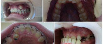
The Popov-Godon phenomenon in the photo:
The results of the analyzes indicate that such a syndrome rearranges the alveolar bones in dental units left without opposite analogues: the periodontal gap narrows, the routes of collagen fibers change. At the same time, certain dystrophic transformations occur in the pulp.
Such situations happen repeatedly and complicate the process of prosthetics, and therefore orthodontic devices have been created that are designed based on the category of removable or fixed dentures. Through them, teeth devoid of antagonists are allowed into occlusion, along with the synchronous separation of other links.
If there is a noticeable movement of the dental unit, the pulp is removed from it and trimmed within the occlusion curve. Significant exposure of cement provokes its extraction.
If it is necessary to wear the plates 18 hours a day, treatment will last at least six months and depends on the complexity of each individual case. If the effect of orthodontic treatment is less positive, prosthetics are performed.
About 30% of the population, having removed a tooth, often do not even think about restoring it, explaining this by disagreement with damaging neighboring links.
Physiologists, thanks to research, have found that when a tooth is lost, a person does not lose the ability to chew food well. But the body does not tolerate emptiness, and the neighboring teeth, moving, try to hide the gap, causing gaps to appear between themselves.
An even more serious situation is where the dental analogue from the opposite area grows towards the void, interferes with proper chewing and increases the functional load, which contributes to the modification of the bite.
Visual video about dental deformation:
Clinical picture
After extraction, a natural compensatory reaction of the body develops. It leads to quite unpleasant consequences - deformation of the dental arch, impaired chewing function, changes in bite and pathologies of the temporomandibular joint associated with overload of the musculo-ligamentous apparatus.
The more teeth are lost, the faster the accompanying disorders develop:
- change in occlusion and inclination of the dentition;
- the formation of a gum pocket and exposure of the roots of the teeth located adjacent to the socket of the missing unit;
- narrowing of the periodontal space;
- pulp dystrophy and changes in periodontal structure.
All these violations in the future significantly complicate the work of doctors if the patient nevertheless decides to install an implant or prosthesis.
Development mechanism
If a denture or implant is not installed, the adjacent teeth gradually move towards the missing unit. In addition, on the opposite jaw, its antagonist begins to “lengthen,” as it were.
The reason for this phenomenon is changes in the periodontium - the so-called adaptive restructuring caused by the loss of the usual functional load. It entails metabolic and microcirculation disorders in periodontal tissues.
At the beginning of the process, tissue hypertrophy occurs, and in the later stages, tissue atrophy occurs, accompanied by partial periodontal resorption.
This is interesting: Choosing an irrigator for braces on the advice of a specialist
Symptoms
The removal of a tooth or several teeth causes a compensatory reaction on the part of the body, which is expressed in deformation of the dental arch and the jaw itself. Neighboring teeth begin to shift towards the missing one. As a result, the spaces between the remaining teeth increase. Not only the dentition in which the tooth is missing is deformed, but also the dentition of the opposite jaw. With the Popov-Godon phenomenon (syndrome), a significant displacement of the tooth in the opposite jaw occurs. The tooth seems to “grow” from the dental arch and creates a “lock” when the jaws move. This disrupts the chewing function and often disrupts the bite. Often, along with the tooth, the jaw bone is also deformed, which creates additional problems during treatment. In some advanced cases, this can lead to problems with the temporomandibular joint due to overload of the ligaments and articular surfaces.
Simultaneously with tooth displacement, an increase in the alveolar process (dentoalveolar elongation) may occur, which leads to the formation of a gum pocket and exposure of the tooth root. Dentoalveolar elongation occurs due to changes in the structure of the periodontium due to the loss of the usual functional load (adaptive restructuring), in which there is a disruption of metabolic processes and microcirculation in the periodontium, an increase in the size of the alveolar process and the volume of bone tissue. At the beginning of the process of adaptive restructuring of the periodontium, an increase in the volume of tissues formed occurs, while the tooth moves beyond the occlusal plane, and in a later period the atrophic process with the phenomena of periodontal resorption (“atrophy from inactivity”) predominates.
Diagnostics
It is based on a visual examination of the oral cavity using a classification of the phenomenon and a detailed study of radiographic data.
In addition, the dentist makes a differential diagnosis of the Popov-Godon phenomenon with partial edentia by assessing occlusion in a state of physiological rest.
To determine the pathology, a specialist examines the oral cavity and assesses the condition of the lower and upper jaw. To accurately determine the shape, it uses the classification of the Popov-Godon phenomenon. It is with its help that the doctor will be able to more accurately determine the stage of development of the disease.
An important point is differential diagnosis, which helps to separate pathology from partial edentia, which has similar clinical manifestations. For this purpose, the specialist compares the ratio of the dentition to each other in a state of physiological rest. It is also necessary to determine the distance between the teeth in the anterior and lateral projections.
The first parameter is considered the most important, since only when a displacement is detected in the central region can we talk about the development of a pathological process. If clinical manifestations are present, but there are no changes in this area, edentia of varying degrees of severity is most often suspected.
An additional method is an X-ray examination and study of the resulting images. After establishing an accurate diagnosis and the extent of the lesion, the specialist determines a treatment regimen that is individual in each case.
Signs
- Partial edentia.
- Deformation of the occlusal curve.
Supra- or infraocclusal position of the teeth, in which the maxillary elements fall below the occlusal curve, and the mandibular elements move above it. The distance between the chewing surface of the displaced units and the alveolar ridge of the opposite jaw decreases until it completely disappears (the teeth touch the soft tissues). - Increase in the volume of the alveolar process.
- The appearance of gum pockets, periodontal resorption (in the 2nd form of the syndrome).
- Blocking LF movements.
At the same time, the occlusal height is maintained, and the remaining teeth remain intact (normal). In the 1st form of the syndrome, the patient does not complain of discomfort or pain.
Differential diagnosis
Differential diagnosis consists of isolating the Popov-Godon phenomenon from other pathologies that may be mistaken for it.
This is interesting: Incognito braces (Germany) - lingual system made of gold alloy
With the syndrome, teeth that do not meet the opposition of antagonists cross the occlusal plane and invade the “foreign” space , while in other pathologies there is no crossing of the occlusal plane.
The P-G phenomenon is sometimes mistakenly mistaken for a decrease in occlusal height due to tooth abrasion, distal displacement of the MF, or the general absence of antagonists in all teeth.
To establish the true picture, the models installed in the occluder are studied, paying main attention to the position of the occlusal plane, the distance between the defective teeth and the soft tissues of the opposite jaw.
It is important that the models are set in centric relation and the normal height of the lower third of the face is maintained.
Treatment options
To eliminate the symptoms of the pathology and prevent its further progression, specialists use several methods of therapy. The following are considered the most popular:
- grinding;
- disocclusion;
- hardware-surgical treatment;
- surgical intervention.
Grinding is used to treat patients over 35 years of age at the initial and middle stages of the disease. Most often it is used in the second form of pathology when the teeth are displaced by no more than half of their own height.
After examining the patient's x-rays, the specialist determines the degree of grinding. The procedure is simple and involves grinding down teeth. If it only removes a small area, after the manipulation the patient undergoes treatment to restore tooth enamel. In cases where the procedure exposes dentin, covering with a crown no later than 2 days after the procedure is considered a prerequisite.
Typically, such treatment does not require much time, and if immediately after it all teeth are restored and implants are placed in the place where it is necessary, the risk of relapse is significantly reduced.
Disocclusion technique
This method brings results only when treating patients no older than 40 years old who suffer from the first form of pathology. It consists of a mechanical effect on crooked teeth using a special device. It is made of special high-strength materials. The first stage of therapy is the introduction of the structure into the oral cavity and comfortable placement.
The device should correct the patient’s bite, straighten the teeth and prevent further progression of the disease. The time required to achieve maximum effect varies depending on the extent of the lesion and the age of the patient. It was noted that in patients under 30 years of age, the alignment process occurs much faster.
In severe cases of protrusion, the design will not help to completely straighten the teeth and correct the bite. In this case, the specialist resorts to the second stage of treatment: covering the upper part of the teeth with a special plastic that quickly hardens. It allows you to separate the teeth and maintain a distance of no more than 2 mm between them. Thanks to this, the bite is corrected, the upper and lower dentition is aligned. This technique is used until the defect is completely eliminated.
After eliminating the violation, the specialist strongly recommends that the patient fill the voids in the oral cavity by introducing implants or dentures. If this is not done, the pathology will begin to develop again and repeated treatment will be longer and more difficult.
Hardware-surgical therapy
A similar technique is used in cases where disocclusion did not bring results, and the gums around each displaced tooth became inflamed. This therapy is especially relevant when the root is exposed and dystrophic changes in the pulp.
The essence of the method is to install special prostheses in the places where the prostheses were extended, which gradually correct the violation. The time it takes for the structure to remain is strictly individual and depends on the degree of dental disease. In medicine, this technique is called compactosteotomy.
Installation of the device requires local anesthesia and special attention in the area of the upper palate. It is in this part that the prosthesis often damages the mucous membranes. After the implementation of the structure, careful care of the oral cavity is required, since the doctor places sutures that need to be processed.
https://youtu.be/Aw8LUcQi0-8
Surgical intervention
The technique is indicated for patients with the second form of the pathological condition, as well as with rapid progression of the disease, the presence of symptoms of gum inflammation, exposure of the tooth root and malocclusion. The procedure is to extract all units that deviate. The operation is performed under general anesthesia.
Before it is carried out, a complete examination of the patient is carried out. The following are considered mandatory steps: laboratory blood test with determination of platelet count and erythrocyte sedimentation rate, as well as glucose level, electrocardiogram, x-ray of the skull with special attention to the jaw. After receiving all the results, the specialist determines the drug that will be used to relieve pain.
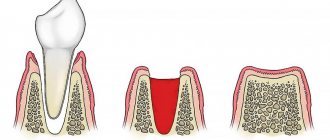
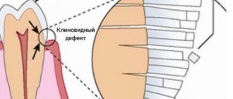
Displacement and exposure of teeth as a result of loss of neighboring
This treatment is strictly contraindicated for patients with persistent arterial hypertension, congestive heart failure, during the recovery period after an ischemic stroke and myocardial infarction, severe bronchial asthma, thrombophlebitis, type 1 diabetes mellitus with frequent attacks of hyperglycemia, bleeding disorders, acute anemia at an advanced stage .
After the operation, the patient may experience severe pain at the site of the extracted teeth, hyperthermia, even fever, weakness, fatigue, nausea and lack of appetite. For several days after the intervention, you are not allowed to eat solid food, as injury to damaged gums may occur.
As a rule, negative reactions after surgery are associated with infection, individual characteristics of the body, or the negative effect of the anesthetic on the central nervous system. To eliminate them, symptomatic therapy is used.
After partial healing, the patient visits the specialist again to determine the time required for complete recovery. This is required so that the dentist can recommend implants or dentures to the patient that will fill the voids and prevent other teeth from protruding. The introduction of artificial structures is considered a must, especially for young people.
Pathogenesis
Histologically it has been established that in teeth devoid of antagonists, the periodontal gap is significantly narrower than in teeth with antagonists.
Statistical data show that in functioning teeth there is no difference between the width of this gap on the vestibular and lingual sides. At the same time, it has been absolutely reliably established that there is a significant difference in the width of the periodontium between the cervical, middle and apical thirds on both the vestibular and lingual sides.
In the periodontal tissue of teeth devoid of antagonists, the number of fibrous bundles is less than in teeth with antagonists, and the fibrous bundles themselves are less powerful. The predominant direction of such bundles in teeth devoid of antagonists is more oblique than in the control, or longitudinal. In the compact plate of the alveolar walls facing the periodontium, and in the ridge of the alveolar walls of teeth without antagonists, the fibrous bone has greater layering than in teeth with antagonists.
Spongiosa in teeth devoid of antagonists is constructed primarily from thinned bone beams that are in the process of restructuring. In teeth that have antagonists, spongiosis is formed by powerful beams located radially in relation to the root.
In the first form of dentoalveolar deformation, the shape of the periodontal gap is preserved in the cervical part of the root, like that of a functioning tooth, but its size decreases, and in the apical part it changes and becomes equal in size and shape to the periodontal gap in the middle part of the root, i.e., it narrows.
In the second form of dentoalveolar deformation, the shape of the periodontal fissure is preserved in the cervical and apical parts of the root on the palatal side, and its value is less than in functioning teeth and greater than in the first form of deformation. On the vestibular side, the shape of the gap changes, and its dimensions in the middle and apical parts are equal. Comparing the size and shape of the periodontal fissure, it can be assumed that in teeth devoid of antagonists, the amplitude of movement in the alveoli significantly decreases and the direction of their movement changes. If we compare the morphological data for deformities of the first and second forms, we can be convinced that with the second form of deformation the periodontal gap is wider. The processes of new formation of fibrous bone in the compact part of the alveolar wall predominate in the first form of deformation. The processes of restructuring of the spongy substance, characterized by thinning of the bone beams and a change in their location compared to the norm, are expressed to varying degrees in deformation of both forms. Thus, with deformation of the second form, the thinning of the bone beams reaches a greater magnitude and is found in a larger number of bone beams.
It can be assumed that the types of deformities observed in the clinic are based on a single process of bone restructuring as a result of the loss of its normal functional load. The structure of the periodontium changes according to new functional conditions, and when a tooth is deprived of antagonists and falls into other functional conditions, the exchange and morphological relationships between the surrounding tissues are disrupted due to a change in function. This restructuring of periodontal tissue is adaptive in nature.
Histological studies of blocks with deformation of the first form (without exposure of the root) showed that, despite the increase in the alveolar process, there is no addition of bone substance, but only the construction of new, thinner bone beams, which is characteristic of teeth devoid of antagonists. This restructuring of the alveolar process with the rearrangement of the bone beams leads to a visible increase in its volume.
Adaptive restructuring of the dental system as a result of long-term reduced function determines the predominance of atrophic processes in the underutilized section.
Based on observations of various manifestations of deformation of the dentition as a result of the absence of antagonists, it can be established that the initial period of adaptation is expressed by the restructuring of bone tissue, especially in the apical region - an increase in newly formed tissue and displacement of the tooth beyond the occlusal plane. The later period is characterized by the predominance of the atrophic process, which is clinically manifested by the exposure of the neck and root of the displaced tooth, and the beginning of the atrophy process in the form of resorption of the ridges of the alveolar walls is histologically determined even with the deformation of the first form. In other words, the first form of the phenomenon gradually transforms into the second over time, and therefore, the clinical forms of manifestation are stages of adaptive restructuring of bone tissue to change the functional load.
Histological studies of corpses of people with deformations of the dentition after the loss of antagonists, as well as experimental observations of the dynamics of bone tissue restructuring in an underloaded link, revealed a picture of adaptive restructuring under changed functional conditions in the dentofacial system.
Early reorganization of bone tissue, which underlies the deformation of the dentition, manifests itself and is accompanied by changes in metabolism. To determine early biochemical changes in jaws whose teeth have partially lost antagonists, an experimental study was conducted using radioactive tracers. A greater amount of radioactive calcium inclusion was determined on that side of the jaw where the teeth were devoid of antagonists.
It is known that the penetration of phosphorus into calcified tissue occurs the faster, the less mineralized the tissue is. Therefore, it can be assumed that radioactive calcium penetrates faster into less mineralized tissue.
Changes in calcium metabolism in an underused area of the dentition are detected by a biochemical method at an earlier time than tissue changes observed during histological examination. Functional load is the most important physiological stimulus that maintains normal mineral metabolism and the histological structure of bone tissue. Therefore, any deviation, either in the direction of increasing or decreasing the mechanical pressure on the teeth, is quickly reflected in the dynamics of the biochemical process occurring in the bone tissue of the jaws. Starting from three months after the loss of antagonists, periodontal fatty degeneration was detected, especially pronounced in cells located near the cementum of teeth devoid of antagonists.
Changed conditions after the loss of some antagonists first cause shifts in the metabolic processes of the jaws, and subsequently lead to morphological changes in both dental and periodontal tissues.
The electrical excitability of teeth devoid of antagonists is reduced - ranging from 12 to 300 μA. The decrease was more pronounced the longer the period from the moment of loss of the antagonists.
An experimental study of the nature of changes in the nerve elements of the jaws during the formation of deformation showed that in the first 3 months, along with the normal structure, hyperargeria and vacuolization were observed in some nerve fibers. In the bone marrow spaces, not only vacuolization was observed, but also fragmentation of individual nerve fibers. After 4-6 months, dystrophic changes in the nervous elements of the periodontium manifested themselves in the form of hyperargeria and cup-shaped thickening, vacuolization and fragmentation. On the part of the bone marrow spaces, a pronounced disintegration of nerve fibers into fragments and their vacuolization was observed.
After 11-12 months after removal of the antagonists, degenerative changes in the nerve fibers of the periodontium and bone marrow spaces were more significant. In the bundle of periodontal nerve fibers, decay and swelling were observed in almost all fibers. Only a few nerve fibers had a normal structure.
These histological studies and studies of metabolic processes indicate the presence of vicarious (adaptive) processes occurring in periodontal tissues in response to the elimination of chewing pressure on the tooth and its periodontal tissues. The term “atrophy from inactivity” is also used in these cases - it develops as a result of a long-term decrease in the functional load on the teeth.
Tensometric studies have established that during the chewing process, the bone tissue of the jaws is under double influence: the general functional stress state and the functional stress in the walls of the alveoli.
Thus, when the second molar is loaded, elastic deformation occurs in its periodontium. The same deformation, but of a different sign and smaller magnitude, is observed in the group of molars on the opposite side. The distribution of deformation is also observed in the presence of defects in the dentition. In the area of teeth devoid of antagonists, general functional tension is maintained and there is no tension from local influence. This, in turn, leads to a change in the nature of the deformation: instead of cyclical alternating deformations of compression and tension, mainly tensile deformation remains. This may explain not only the change in the position of the bone crossbars, but also the predominance of the resorption process over the process of bone tissue formation.
The study of elastic deformations of the bone tissue of the jaws makes it possible to confirm and clarify the mechanism of tooth movement described by Godon during the loss of part of the teeth, including the loss of antagonists.
Based on the correct position that the dental system is a single whole, Hodon considered the possibility of the existence of stability of this system while maintaining the continuity of the dentition. Each tooth has contact with the adjacent one and when loaded, its pressure is transmitted throughout the entire dentition. Hodon presented the relationship of the tooth with the antagonist and the one standing next to it at the moment of functional load in the form of a parallelogram of forces. When even one tooth is lost, the direction of forces on the teeth bordering the defect changes. Thus, when an antagonist is lost, the tooth does not experience any load coming from it, and the action of forces from neighboring teeth seems to stimulate movement beyond the occlusal curve. The absence of direct load and the action of lateral forces stimulate the process of bone formation in the periapical region and a change in the course of the bone trabeculae.
The processes of bone tissue restructuring, as a rule, proceed slowly, since general functional elastic deformations are retained during chewing and an adaptive reaction develops aimed at preserving periodontal function. Over time, the adaptive reaction fails, which is manifested by the development of the second form of the Popov-Godon phenomenon.
Tartar deposition also plays a significant role in the pathogenesis of the phenomenon, since the process of tooth cleansing is difficult due to the lack of movement of the food bolus. Tartar deposition aggravates the process of resorption of marginal periodontal tissues.
As a result of edentia and the severity of the phenomenon, depending on the time of tooth loss and their number, a change occurs in the muscular system, manifested primarily by an asymmetry of tone and contractile force.
On the deformation side, the function of the masticatory muscles changes. These changes are expressed in a weakening of muscle tension and an increase in the chewing period. On myograms this is manifested by a sharp decrease in the amplitude of the waves and a change in their character. A small amplitude of the teeth indicates weak muscle tension. Steep ascents and descents of curves reflect rapid changes in muscle tension. The difference in the nature of the teeth reflects arrhythmic muscle tension. The chewing time of an almond kernel is extended from 37 to 63 seconds.
Disease prevention
The most effective method of preventing a pathological condition is considered to be the prevention of the development of dental diseases that require their removal. Oral hygiene and avoiding injuries or permanent mechanical damage will help prevent such diseases.
Despite following all the rules, tooth loss is inevitable due to age or certain pathologies associated with calcium metabolism disorders. They lead to the gradual destruction of enamel, crown, and dentin. In cases where removal cannot be avoided, it is necessary to quickly replace it with a prosthesis or titanium implant, which will fill the voids and prevent displacement and elongation of the alveolar processes.
The Popov-Godon phenomenon is a severe dental pathology, accompanied by pronounced clinical manifestations and interfering with the normal functioning of the teeth. In the absence of therapy for a long period, the symptoms can only be eliminated through surgery and implantation.
Preventive actions
The development of the phenomenon will help to avoid simple measures:
- Pay attention to oral hygiene.
- Prevention of diseases leading to tooth loss.
- Immediate replacement of a lost organ with a prosthesis or implant.
It is easier to prevent any disease than to treat it later. It’s impossible to avoid everything, but everyone can visit a dentist in a timely manner.
The video provides additional information on the topic of the article.