The main function of the salivary glands is the formation of a secretion that helps soften food, form a food bolus and facilitate its movement through the esophagus.
A whole complex of glands is responsible for the formation of saliva in the human oral cavity:
- three pairs of large ones
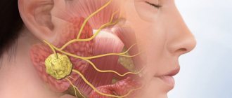
: parotid (located just below the auricle), submandibular (located under the lower jaw) and sublingual (located on both sides of the tongue); - many small ones
, divided by location into molar, labial, lingual, buccal and palatal.
In medical practice, there are various pathological conditions of the salivary glands that affect their functionality and cause a number of unpleasant symptoms. Patients of different age groups are diagnosed with: sialadenitis, mumps, duct blockage, salivary stone disease, benign and malignant tumors.
The provoking factors contributing to the development of the inflammatory process are:
- infectious diseases: influenza, pneumonia, encephalitis, etc.;
- disturbances in the functioning of the endocrine system;
- injuries and surgical interventions;
- taking medications that reduce saliva production and thickening;
- dehydration and malnutrition.
Each disease produces its own set of symptoms, the severity of which depends on the nature of the inflammation and the location of the affected gland.
Symptoms of sialadenitis
With the normal functioning of all glandular components, saliva is formed in sufficient quantities in the oral cavity, and its composition is optimal for digestion. If sialadenitis occurs, symptoms such as:
- change in the amount and consistency of saliva;
- change in taste sensations;
- limited jaw movements;
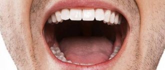
- pain while eating;
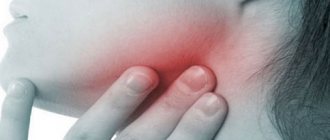
- swelling or even severe swelling in the area where the inflamed gland is located.
With an acute inflammatory process or the development of an abscess, an increase in body temperature and a sharp deterioration in general health are also observed.
In the initial stage of the disease, a person may not notice the manifestations of the disease or not pay attention to the symptom until the inflammatory process causes pain and swelling.
Important! Inflammatory processes in the salivary gland tend to recur frequently and develop into a chronic form. Therefore, even if the condition has improved sharply soon after the onset of the disease, it is necessary to be examined and undergo a course of treatment recommended by a specialist.
Symptoms of sialolithiasis. Causes of the disease and how to treat it
come back
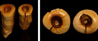
Sialolithiasis (or salivary stone disease) is a disease that is accompanied by inflammation and the formation of stones in the ducts of the salivary glands.
The appearance of salivary stones (calculi) is caused by stagnant processes in the glands. Most often, the pathology affects the submandibular gland, much less often the parotid gland and, in rare cases, the sublingual salivary gland.
In most cases, sialolithiasis develops between the ages of 20 and 40 years. Moreover, men are more susceptible to it than women. And salivary stones found in patients consist of deposits of inorganic salts - calcium carbonate or phosphate. In rare cases, they contain iron, potassium, magnesium and sodium. When the process is advanced, the stones can reach the size of a chicken egg.
Causes of the disease
The direct causes of sialolithiasis have not yet been determined by science. However, there are a number of factors that can trigger the development of the pathological process. These include:
- disturbances in the functioning of the salivary glands;
- previous injuries to the glands;
- stagnation of salivary fluid due to individual characteristics of the jaw structure;
- disturbance of calcium metabolism in the body;
- vitamin A imbalance;
- increased blood clotting;
- the presence of fungal and infectious diseases - mumps, tuberculosis, syphilis.
Often the provoking factor is the presence of a foreign body in the salivary duct. A piece of toothbrush bristles, a crumb of cracker or a salt crystal in this case acts as a kind of “magnet” that attracts inorganic salts that form salivary stone.
Symptoms of sialolithiasis
The initial stage of the disease is characterized by a complete absence of symptoms. As the stone forms and increases in size, tingling pain and slight swelling in the area of the salivary glands may be present.
Further progression of the pathological process causes the appearance of pain, which intensifies during meals. In this case, the patient experiences a sharp increase in body temperature and a deterioration in general health. A particularly striking clinical picture is observed in the case of movement of salivary stones into the oral cavity.
Other signs of the disease include the following symptoms:
- presence of dryness and unpleasant taste in the mouth;
- bursting pain that intensifies during swallowing, opening the mouth and normal conversation;
- mild or severe swelling of the salivary glands;
- general weakness, chills and headaches.
Important to remember! Attentive attention to your own well-being, timely detection of the first symptoms of the disease and seeking help guarantee a successful cure for the disease.
Diagnosis of the disease
If during the initial examination the patient is found to have pain, a dense structure and an increase in the normal size of the salivary glands, then he is prescribed additional tests.
- Ultrasound, which helps to identify formed stones and their location.
- Computed sialotomography is a method that helps determine the presence and location of X-ray negative salivary stones.
- Contrast sialography, with which you can get a clear picture of the structure of the salivary ducts.
- Biochemical analysis of salivary fluid, thanks to which it is possible to establish the composition of inorganic salts that formed the stone and differentiate them from other diseases.
Additional research methods are prescribed by the attending physician.
Treatment of sialolithiasis
The choice of treatment methods directly depends on the stage of formation of salivary stones and the presence of exacerbation of the pathological process. Therapy includes surgery and the use of medications.
- Surgical intervention is carried out both at the stage of exacerbation of the process and during the chronic course of the disease. After removal of the stones, treatment is provided aimed at relieving the inflammation process and restoring the functions of the gland.
- Drug treatment involves the use of antibiotics and drugs that dissolve stones.
If during the study it was determined that the stones cannot be removed, then specialists carry out complete removal of the salivary gland along with the stone (extirpation).
Folk remedies for treating the disease</>
To treat sialolithiasis, traditional medicine suggests using the following methods.
- Pharmacy tincture of echinacea, which is diluted with boiled water at room temperature in a 1:1 ratio and used for compresses.
- A solution of baking soda, for which 1 tablespoon of soda is dissolved in 1 glass of warm boiled water and used for rinsing the mouth.
- Homemade ointment, for the preparation of which you mix 1 ampoule of Novocaine, 1 raw chicken egg, 1 teaspoon of honey and olive oil. The resulting mixture is applied to the inflamed areas 3 times a day for 7 days.
Important to remember! The use of traditional medicine is possible only at the initial stage of the inflammatory process! While slightly alleviating the manifestation of pain, it does not replace surgical intervention and drug therapy, for which it is necessary to contact specialized specialists. Otherwise, the development of the disease threatens a number of complications.
Possible complications of sialolithiasis
Untimely contact with specialists can provoke the development of complications of the disease - abscess of the soft tissues surrounding the area where the stone is located and the formation of external fistulas.
The prognosis of the disease, subject to timely and adequate treatment, is favorable.
Disease prevention
All preventive measures are aimed at eliminating possible factors that can cause the formation of salivary stones. These include:
- maintaining careful oral hygiene;
- rejection of bad habits;
- balanced diet rich in vitamins;
- timely treatment of fungal and infectious diseases.
If the first signs of sialolithiasis appear, you should immediately seek help from specialized specialists - a dentist and a surgeon.
Symptoms of mumps
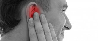
Mumps is an inflammation of the parotid gland caused by a viral infection. This pathology is characterized by an acute onset of the disease. Parotid swelling occurs, painful when palpated, and a high temperature rises.
There is no specific treatment for this disease. Therapy comes down to relieving symptoms and providing comfort to the sick person.
The main dangers of mumps are its serious complications:
- acute pancreatitis;
- orchitis;
- oophoritis;
- ovarianitis;
- meningoencephalitis.
Reliable insurance against such serious consequences as the development of infertility in men and women, diabetes mellitus or deafness is vaccination.
Why does inflammation of the salivary glands begin?
Inflammation of the salivary glands occurs in two types.
Epidemic sialadenitis
The causes of epidemic sialadenitis are viral diseases and infections. A common cause of inflammation of the salivary glands is mumps. The virus in this disease passes from one person to another through airborne droplets. The inflammatory process in the salivary glands occurs with a strong increase in their size.
Non-epidemic sialadenitis
Another cause of inflammation of the salivary glands is the formation of a blockage of the salivary duct. Disturbances in the functioning of the duct can occur due to severe mechanical injuries, the entry of foreign bodies into them, or the development of salivary stone disease in a person - sialolithiasis.
A very common reason for the development of inflammation of the salivary glands is irregular adherence to oral hygiene rules. Non-epidemic parotitis - this is the medical name for inflammation of the parotid salivary glands - can occur as a result of an infectious disease (pneumonia, influenza, typhus, encephalitis are at risk) or as a consequence of surgical intervention.
Tumors of the salivary glands
About 60% of all diagnosed neoplasms of the salivary glands are benign in nature. These are adenomas, adenolymphomas, papillary cystadenolymphomas and polymorphic tumors. The remaining 40% are intermediate stages (mucoepidermoid neoplasms and carcinomas), as well as sarcomas and cancers, accounting for 10-25% of cases.
To make a correct diagnosis, a complete examination is necessary, which includes, in addition to the standard set of laboratory tests, diagnostic methods such as:
- Ultrasound;
- X-ray examination;
- MRI;
- biopsy;
- histological examination.
In most cases, surgical treatment is indicated, which consists of removing the tumor node.
Sialography
The most common and informative method for diagnosing the salivary glands, which allows us to identify structural changes that are fundamentally important for making a diagnosis that have occurred in the gland and ducts. Sialography also helps to identify the presence and exact location of stones, diagnose cysts and tumors of the salivary glands. The method consists of conducting an X-ray examination with contrast of the salivary glands.
Sialometry serves to assess the secretory function of the salivary glands and determine the degree of xerostomia. For its behavior, a special cannula is installed at the mouth of the excretory duct and salivary secretion is collected for 5 minutes, while salivation is stimulated.
Local treatment consists of bougienage and instillation of the ducts of the salivary glands, novocaine blockades, prescription of medications, and for salivary stone disease - removal of stones.
The principle of treatment of sialadenitis is aimed at stopping the inflammatory process, eliminating the etiological factor, treating general somatic pathology and dynamic observation.
Which doctor should I contact for help?
Sometimes a dentist can diagnose sialadenitis, which occurs without pronounced symptoms, when examining the oral cavity. But, in the case of a pronounced tumor, which is accompanied by pain and fever, it is worth contacting a therapist as soon as possible, who will perform an examination, prescribe the necessary diagnostics and recommend which doctor to make an appointment with. For uncomplicated inflammation, conservative treatment will be effective, including:
- taking medications (antipyretic, painkillers, and, if necessary, antibacterial or antiviral);
- compresses;
- physiotherapeutic procedures.
In some cases, consultation with a surgeon, endocrinologist or gastroenterologist may be required, depending on the complications that arise.
Consultations with a doctor online Taking care of your health is a life priority for everyone. Communicate with doctors online and receive qualified assistance without leaving your home. Try it
Note! The information on this page is provided for informational purposes only. To prescribe treatment, you must consult a doctor.
Examination of the thymus gland
The thymus or thymus gland is an organ located behind the sternum. It ensures the normal development of the body's protective functions. When an infection enters a child’s body, the thymus participates in the formation of immune system cells (T-lymphocytes). The growth of the thymus continues up to 12 years. At this age, the child’s immune system is finally formed and the growth of the thymus stops. With age, gradually, the thymus gland atrophies and decreases in size. Usually the reason for examination is an enlargement of the thymus gland: thymomegaly, thymoma. In this case, it is important to understand the reasons for the enlargement of the thymus and offer the optimal solution: removal of the thymus or treatment without surgery. Indications for surgery may include:
- Myasthenia gravis
- Compression of adjacent organs by the enlarged thymus gland, if this significantly impairs their function.
We can offer basic research methods that are used in the diagnosis of thymus diseases:
- Ultrasound of the thymus is a fast, safe and simple method for examining the thymus. But visualization of the thymus using ultrasound is hampered by the bones of the chest; ultrasound does not provide sufficient visualization of the intrathoracic lymph nodes, determine the degree of pressure of the thymus on nearby organs (esophagus, mediastinum, lungs), etc. Therefore, CT – computed tomography of the chest organs – is preferable. MRI is less commonly used.
- MRI and CT scan of the thymus - on tomograms you can see the smallest details in the structure of the thymus gland and even recreate 3D images of the thymus.
- If we are talking about immunodeficiency or autoimmune pathology, then it is advisable to perform an immunogram and conduct an examination for infections that can cause enlargement of the thymus. These are, first of all, infections of the herpes group. To do this, we will offer you to donate blood and saliva for PCR testing and determination of antibodies in the blood to herpes viruses (herpes types 1, 2, 6, Epstein-Barr virus, cytomegalovirus).
- If we are talking about myasthenia gravis, i.e. autoimmune complication of thymoma (tumor of the thymus), we recommend that you perform a study of antibodies to acetylcholine receptors and antibodies to skeletal muscles. Also in this case, electromyography (needle, decrement test).
In rare cases, enlargement of the thymus may be caused by lymphoproliferative diseases or more severe infections, for example, brucellosis, chlamydial infection, mycoplasma, etc. in conditions of impaired immune response.
Diagnosis and treatment of inflammation of the salivary glands
How is sialadenitis diagnosed?
Sialadenitis is diagnosed by a dentist during an examination of the oral cavity or by a therapist if you go to a multidisciplinary clinic with the disease. During the examination, a significant increase in the salivary glands is usually detected, and sometimes the discharge of purulent fluid. In the case of a bacterial infection, the salivary glands become painful.
If the attending physician suspects an abscess, the patient is usually prescribed an ultrasound examination. Our Dentistry is located in the same building as the multidisciplinary clinic “Diamed” on Shchelkovskaya. You will be able to quickly receive treatment and undergo the necessary diagnostic and treatment procedures.