Salivary gland cyst
Patients are concerned about a round-shaped formation, small at first, then slowly increasing, not causing pain. Sometimes, when injured by food, it is emptied, then it fills up again. Objectively: a round-shaped formation is determined under the mucous membrane of the lower lip, cheek or in another location; usually the mucous membrane above it is not changed. As the secretion accumulates, the color of the mucous membrane may acquire a blue tint; upon palpation, the consistency of the formation is soft-elastic and moves freely.
Differential diagnosis is made with hemangioma (with hemangioma, after pressing, the formation disappears, if the pressure stops, it fills again).
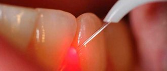
Surgical treatment: under local anesthesia, two bordering incisions are made in the mucous membrane above the surface of the cyst, then it is peeled off, holding the edges of the mucous membrane, and the wound is sutured with catgut.
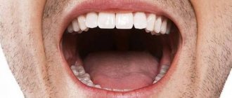
A cyst of the sublingual salivary gland (ranula) is most often located in the sublingual area above the mylohyoid muscle and resembles a bubble filled with fluid. With large sizes, it can shift the frenulum of the tongue to the other side. Less commonly, the cyst penetrates into the submandibular region and macroscopically looks like an hourglass, located in the supra- and sub-hyoid muscle, narrowing at the site of its perforation.
Patients complain of a formation under the tongue, which slowly increases, beginning to interfere with eating and talking. It can be periodically emptied and then refilled.
Upon examination, an oval-shaped formation is detected in the sublingual area, which, if large, can spread to the opposite side. The mucous membrane above it becomes thinner and underneath it it is possible to identify a cavity filled with transparent contents. On palpation, the formation has a soft-elastic consistency, limited from the surrounding tissues by the capsule. Differential diagnosis should be made with a dermoid cyst, salivary stone disease, submandibular salivary gland cyst, lipoma, sialadenitis.
Rarely, a cyst of the sublingual salivary gland becomes infected and then it must be differentiated with an exacerbation of chronic sialadenitis and salivary stone disease with localization of the salivary stone in the excretory ducts. To clarify the diagnosis, a puncture can be performed: a viscous mucous fluid will be obtained from the cyst. To exclude salivary stone disease, a survey radiography is performed. Cystography can be used to diagnose a cyst.
Treatment is surgical. If the cyst is located above the mylohyoid muscle, then the most radical way is to remove the cyst along with the gland. However, its use is limited due to the fact that the cyst shell is very thin and easily damaged. After which the cyst is emptied, the walls of the cyst collapse and it can be very difficult to separate the cyst shell from the underlying tissues.
Therefore, the method of cystostomy proposed by I. G. Lukomsky (1943) has not lost its significance to this day. Under local anesthesia, the protruding part of the mucous membrane and the upper wall of the cyst are excised, the edges of the mucous membrane and the remaining cyst membrane are sutured around the perimeter, an iodoform tampon is loosely placed on the bottom and fixed by tying the ends of the suture material above it. The tampon is changed after 5 days.
If the cyst spreads to the submandibular region, then the operation is performed in two stages (Kabakov B.D., 1978). First, in the submandibular region, retreating 2.0 cm, and parallel to the edge of the lower jaw, an incision is made in the skin with subcutaneous fatty tissue and superficial fascia, the maximally bulging part of the cyst is isolated to the point of narrowing, it is bandaged at this level and cut off, the wound is sutured in layers, leaving a rubber graduate. Then, in the second stage, the sublingual salivary gland with the cyst is removed or a cystostomy-type operation is performed.
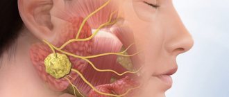
A cyst of the parotid salivary gland appears for no apparent reason; facial asymmetry is clinically determined due to swelling of the soft tissues of the parotid region, which gradually increases, the skin on it is not changed. Upon palpation, a formation is determined to be round in shape, soft-elastic consistency, delimited from the surrounding tissues by a membrane, mobile, no pain.
Differential diagnosis is carried out with chronic lymphadenitis and benign tumors. You can use ultrasound, puncture, sialography in combination with cystography (double contrast).
Surgical treatment: the cyst is removed within the membrane with the adjacent tissues of the salivary gland, the branches of the facial nerve are preserved.
A cyst of the submandibular salivary gland is rare; there is an enlargement of the submandibular salivary gland, slowly progressing. By palpation it is sometimes possible to identify a round formation with a soft elastic consistency. Differential diagnosis is carried out with chronic submandibulitis, lymphadenitis, and benign tumors. During puncture, a yellowish liquid with a viscous consistency is obtained, ultrasound is used, and sometimes cystography is performed.
Surgical treatment: the cyst is removed along with the gland. Cysts of the minor salivary glands are more common, and cysts of the sublingual salivary glands are somewhat less common. Parotid and submandibular salivary gland cysts are rare.
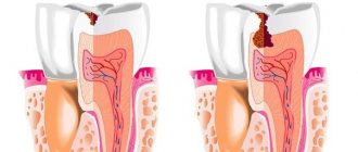
It is believed that cysts appear as a result of retention of the excretory duct, as a consequence of its injury or inflammatory process in the salivary gland and adjacent tissues. There is also a theory that cysts are congenital in origin.
Minor salivary gland cysts most often occur in the lower lip area. The cyst has a connective tissue capsule; the contents of the cyst are a viscous translucent liquid, reminiscent of stagnant saliva.
Publications in the media
Skin papillomas - see Warts, Human papillomavirus infection. Trichoepithelioma multiple familial (*601606, 16q12–q13 and 9p21, MFT, TEM, Â genes) is a benign skin tumor originating from hair follicles, a relatively common inherited dermatosis with many small tumors, more often on the face.
Cysts are fluid-filled cavities • Epidermal cysts; treatment - excision • Sebaceous cysts are the result of blockage of the excretory ducts of the sebaceous glands. Treatment is excision (prevents recurrence). For infected cysts, they are limited to opening and draining the abscess with subsequent excision in a planned manner (they often recur when part of the capsule is left) • Dermoid cysts arise as a result of a violation of embryogenesis; when localized in the midline (glabella, nose), it is necessary to exclude communication of the cyst with the cranial cavity (cerebral hernia); treatment - excision • Synovial cyst - a cyst with fibrous walls containing a thick clear fluid rich in mucopolysaccharides; usually associated with underlying tendons; usual localization - palms and feet; Treatment is excision; with incomplete resection of the pedicle and walls of the cyst, relapse is possible.
ICD-10. L72.0 Epidermal cyst.
Vascular birthmarks are classified according to location and cellular composition. • Hemangioma (strawberry spot) - a soft red formation with a raised surface; characterized by the presence of proliferating mast cells and rapid growth during the first year of life; localization - head, neck, torso and limbs. Spontaneous recovery is possible; Surgery or steroid therapy is indicated for hemangiomas that cause functional impairment (if localized in the eyes, ears, or pharynx).
• Vascular malformations are divided into capillary, venous and lymphatic •• Capillary hemangiomas (“port wine stains”) are most often localized on the face, chest and limbs, observed in Sturge-Weber and Klippel-Trenaunay-Weber syndromes. These are clusters of dilated capillaries in the papillary, dermal and subdermal layers. For small tumors, the method of choice is excision. Laser therapy and sclerotherapy are also effective. • Venous malformations (cavernous hemangiomas) often affect deep structures, including muscles. Platelet sequestration is possible. Treatment is excision, possibly introducing a sclerosing agent into the cavity. •• Lymphatic malformations (lymphangiomas, cystic hygromas) cause hypertrophy of the affected soft tissues. Treatment is excision. A common complication is seroma. •• Arteriovenous aneurysms can suddenly increase in volume, causing compression of surrounding tissue. The treatment method is excision.
ICD-10. Q82.5 Congenital non-neoplastic nevus
Vascular tumors • Pyogenic granuloma (bothryomycomoma) is a tumor-like formation of red or brown skin on a stalk, which is a proliferation of granulation tissue with a large number of capillaries, localized on the face, chest and fingers, and can bleed. Surgical excision or cryodestruction is indicated • Stellate nevi (telangiectasia) occur at any age on the face, chest and limbs. They consist of a central arteriole with radiating vessels similar to venules. An increase in nevi is possible during pregnancy and liver failure. Bleeding is rare. Treatment: laser destruction, electrocoagulation, cryotherapy, sclerotherapy.
Seborrheic keratosis is characterized by light and dark brown papules. If malignancy is suspected, a biopsy is necessary. Treatment is electrocoagulation.
ICD-10. L82 Seborrheic keratosis
Keloids are growths of fibrous tissue in areas of injury and scarring. Treatment is excision. Sometimes auxiliary local GC therapy, electrophoresis or lidase injections, and laser irradiation are indicated.
ICD-10. L91.0 Keloid scar.
Lymphadenitis
An acute nonspecific process manifests itself with pain in regional lymph nodes and an increase in their size. In the catarrhal and hyperplastic form, enlarged nodes can be easily palpated, their pain is insignificant, and general disorders are mild or absent. Lymphadenitis often occurs with the involvement of lymphatic vessels - lymphangitis.
In case of suppuration, the node becomes dense and painful, general intoxication develops - fever, loss of appetite, weakness, headache. Local phenomena increase - hyperemia and swelling in the area of the affected node, the contours of the lymph node become unclear due to periadenitis. The patient is forced to spare the affected area, since the pain intensifies with movement. Quite soon, purulent melting of the lymph node occurs and fluctuation becomes noticeable in the area of infiltration.
If the formed abscess is not opened in time, pus may leak out or into surrounding tissues. In the latter case, adenophlegmon develops, which is characterized by a diffuse, dense and painful infiltrate with individual areas of softening. In the putrefactive form of lymphadenitis, gas crepitus (crunching) is felt when palpating the node. During destructive processes, general disorders progress - fever, tachycardia, and intoxication increase.
Lymphadenitis in children occurs rapidly with high fever, malaise, loss of appetite, and sleep disturbances. Possible serious complications may include generalization of infection with the development of sepsis.
In chronic nonspecific lymphadenitis, the lymph nodes are enlarged, slightly painful, dense, and not fused with the surrounding tissues. The outcome of chronic lymphadenitis is wrinkling of the nodes due to the replacement of lymphoid tissue with connective tissue. Sometimes the proliferation of connective tissue causes a disorder of lymph circulation: edema, lymphostasis, elephantiasis.
Specific gonorrheal lymphadenitis is characterized by enlargement and severe tenderness of the inguinal lymph nodes. Tuberculous lymphadenitis occurs with high fever, severe intoxication, periadenitis, and often necrotic changes in nodes. Lymphadenitis in syphilis is characterized by a unilateral moderate enlargement of the chain of lymph nodes, their lack of adhesion to each other and to the skin. With syphilitic lymphadenitis, suppuration of the lymph nodes never occurs.