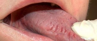
Salivary stone disease (sialolithiasis) is a disease of the salivary glands. Disturbances occur, a malfunction in their work (more often they become inflamed), which leads to the appearance of stones (one or more) in them and their ducts. The presence of stones is always accompanied by inflammation of the glands, but doctors have not yet decided what is primary - stones or inflammation. Typically, such hard formations are found in the submandibular and sublingual glands. They are rare guests in the parotid gland.
In most cases, middle-aged men (30-45 years old) suffer from salivary stone disease. In women, this disease is much less common. It is practically not diagnosed in children.
Causes
Scientists have not fully figured out the exact causes of this disease. Let's get acquainted with their hypotheses. Here are some of them.
- Congenital pathologies of the salivary glands themselves or their ducts (narrowing, wall defects).
- Malfunctions of the salivary glands, changes in the composition of saliva. Much less saliva is produced.
- Infection enters the salivary glands or ducts, resulting in their inflammation.
- Reduced immunity. Disorders of phosphorus-calcium mineral metabolism.
- Lack of vitamins (especially vitamin A).
- Injuries to the salivary glands (tooth crowns).
- Entry of a foreign body (fish bone, toothbrush bristles).
- Increased blood clotting (thick blood promotes the formation of stones).
At risk are patients with a number of diseases (urolithiasis, cholelithiasis, diabetes mellitus).
Sialendoscopy as a method for removing stones from the salivary gland
Chronic obstructive pathologies of the salivary glands can be caused by the presence of salivary stones, the formation of a mucus plug, duct stenosis, the influence of foreign bodies, or anatomical variations in the gland duct system, which can result in retention of saliva in the duct, discomfort, and even infection. Specifically, sialolithiasis is a condition in which a calcified mass forms in the salivary gland. Most often, this disorder is observed in the area of the submandibular gland (in the so-called “Wharton’s duct”) and can cause swelling and pain in the affected area, especially during stimulation of salivation. If the stone does not come out of the gland on its own, it must be removed surgically.
To diagnose such pathologies, methods of radiosialography, ultrasound examination and magnetic resonance sialography can be used. Radiosialography is the most common method for identifying stones in the salivary gland duct, but its use is contraindicated in cases of acute infection or if the patient is hypersensitive to the contrast agent. Ultrasonography is a first-line diagnostic method that allows non-invasively and without the use of contrast to visualize the presence of a calculus in the salivary gland. It should be remembered that the parameters of sensitivity and specificity of this method vary depending on the stage of mineralization of the stone. For diagnostic purposes, of course, you can use more expensive methods such as computed tomography, magnetic resonance imaging or scintigraphy.
Currently, sialoendoscopy is widely used as a non-invasive approach for diagnosing and treating pathologies of the salivary glands. This method allows for direct access to the gland duct, its expansion and irrigation, as well as to establish the presence or absence of patency of the duct.
In this article, we describe the clinical cases of two patients who underwent sialolithotomy procedures using a sialoendoscope at Korea University Anam Hospital. In addition, we will describe the differences between sialoendoscopy and other methods for diagnosing and treating sialolitis.
Clinical case 1
Patient Information
An 81-year-old female patient presented to the dental clinic with a sensation of a foreign body under her tongue and symptoms of dry mouth. She was previously diagnosed with thyroid carcinoma in June 2003 and underwent a total thyroidectomy procedure. The patient's medical history included hypertension and chronic kidney disease. The stone was identified in the duct of the right submandibular gland (Figure 1).
Figure 1. Salivary stones measuring 2 × 10 mm were identified in the right submandibular gland using CBCT.
Surgical procedure
The working area was treated with betadine. After disinfection, the oral cavity was washed generously with saline solution. Local anesthesia was performed in the area of the tongue and floor of the mouth with a 2% lidocaine solution with an epinephrine concentration of 1:100,000. Afterwards, the tongue was sutured with 3-0 silk thread in order to provide optimal conditions for the operation. Using a microscope, the entrance to the duct of the right mandibular gland was found, after which it was entered with probe No. 0000, and then expanded to the size of probe No. 3 (photo 2). In order for the endoscope to penetrate the lumen of the duct, it was expanded using a dilator. The sialoendoscopy procedure was performed under copious irrigation with saline solution. The stone was removed using a three-wire ring (loop) (photo 3). The size of the opening of the duct was smaller than the size of the stone, so the doctor had to make a 0.1 cm incision at the entrance to the gland duct. After removing the stone, we examined the gland with an endoscope to make sure there was patency (photo 4). The glandular system was washed with a steroid solution. No bleeding, swelling or pain was noted in the postoperative period. The patient underwent daily dressings of the intervention area and monitoring for the development of possible wound infection.
Photo 2. a. Gland duct probe. b. Orifice dilator. c. Endoscope monitor. d. Three-portable endoscope. e. Three-wire ring (loop).
Photo 3. a. Stones in the gland duct. b. Removing stones using a loop. c. Capture the stones with a loop and remove them from the duct.
Photo 4. Removal of stones through the mouth of the submandibular salivary gland. b. View of removed stones.
Clinical case 2
Patient Information
A 66-year-old man sought dental attention due to a blocked duct of the right mandibular gland (Figure 5). Approximately 7-8 years ago, he noticed swelling and pain in the lower neck while eating. The sialolithotomy procedure was performed 4-5 years ago in another hospital, but the stones were never completely removed due to their pathological mobility during the operation. The patient had no systemic health problems.
Figure 5. Salivary stones measuring 3 × 4 mm were identified in the right submandibular gland using CBCT.
Surgical procedure
The working area was treated with betadine. After disinfection, the oral cavity was washed generously with saline solution. The patient was given full anesthesia via nasotracheal intubation, and the tongue was sutured with 3-0 silk suture. The intervention protocol was similar to the one we have already described in the first clinical case (photo 6). No bleeding, swelling or pain was noted in the postoperative period. The patient underwent daily dressings of the intervention area and monitoring for the development of possible wound infection.
Photo 6. View of the removed stone.
Discussion
Symptoms of obstructive salivary gland pathologies include recurring, painful swelling of the major salivary glands, which can negatively affect the patient's quality of life. Previously, radiosialography, sonography and MR sialography methods were used to diagnose diseases of the salivary glands. Radiosialography is the main method for examining the salivary glands, which can be used to diagnose sialolithiasis by analyzing images obtained after the injection of contrast into the gland duct. Contraindications to the use of this method are similar patient reactions to the contrast agent and the presence of an acute infectious lesion. Sonography is a non-invasive method for diagnosing stones in the structure of the salivary glands, however, the effectiveness of this approach largely depends on the experience of the doctor conducting the diagnosis. MR sialography allows for complete visualization of the entire salivary gland system, however, the resulting diagnostic images may be characterized by the presence of various types of distortions and artifacts, especially in cases of adjacent localization of dental restorations. However, all of the above-mentioned limitations characteristic of different diagnostic methods can be overcome using the sialoendoscopy method. It is less invasive than other approaches to diagnosing salivary gland pathologies and can be successfully used to assess the condition of their ducts and internal structure.
Several research groups have examined how satisfied patients were with the sialoendoscopy treatment process. Kroll et al, using the short version of the SF-36 questionnaire, found that patients reported a high level of satisfaction after sialoendoscopy. Aubin-Pouliot et al, using a questionnaire designed to assess chronic obstructive sialadenitis, demonstrated similar results. In addition, an interesting fact was established that after sialoendoscopy on the submandibular gland, the symptoms decreased much more pronounced than after a similar manipulation on the parotid salivary gland.
Recent studies have shown that microsialoliths play an important role in the pathogenesis of chronic sialadenitis. They can accumulate in normal salivary glands and provoke the development of obstructive atrophy. The latter, in turn, promotes the colonization and proliferation of microbes, causing inflammation in the peripheral ductal system, accompanied by even more severe atrophy and progressive infection, leading to chronic sialadenitis. According to Quinn et al, intraductal placement of antibiotics facilitates their penetration into the parenchyma of the glands, allowing the existing symptoms of the lesion to be completely eliminated. However, the same results were obtained when irrigating the internal structure of the glands with saline solution.
Radioactive iodine (RAI) therapy is another cause of salivary gland disease. According to Kim, chronic sialadenitis is the most common complication of RAI, especially in cases after thyroidectomy. The prevalence of chronic sialadenitis associated with RAI is 11–65%. Damage to the salivary glands caused by radioactive iodine leads to the development of obstructive sialadenitis and recurrent swelling with or without pain during meals. This chronic condition subsequently causes hyposalivation and associated symptoms, such as difficulty swallowing, taste disturbances, oral candidiasis and caries. Currently, chronic sialadenitis caused by radioactive iodine therapy is treated conservatively by maintaining a good level of oral hygiene, frequent hydration, the use of saliva substitutes, and stimulation of the salivary glands.
According to Kim's study, sialoendoscopy demonstrates similar effects to sialocentesis (intraductal irrigation with sterilized saline) and can improve obstruction symptoms as early as 3 months after mechanical dilatation with an endoscope. However, in patients with chronic sialoadenitis caused by radioactive iodine therapy, sialoendoscopy has been found to have limited ability to relieve existing symptoms of xerostomia.
This article described examples of using the sialoendoscopy method to remove stones from the salivary glands. This method is more effective compared to other approaches to the treatment of sialolithiasis, and at the same time less uncomfortable. In the two clinical cases analyzed, patients did not demonstrate any significant complications or similar effects after performing the manipulation.
Conclusion
This article describes two clinical comparisons of non-invasive sialolithotomy procedures using sialoendoscopy. This approach can be effectively used not only to treat cases of sialolithiasis, but also for ductal stenosis and sialadenitis. At the same time, sialoendoscopy is a minimally uncomfortable procedure, which is characterized by a low level of complications. To formulate any definite recommendations regarding the use of sialoendoscopy in cases of stones in the salivary glands, it is necessary to ensure that studies are carried out with the participation of a larger number of subjects.
Authors: Dong-Keon Lee, Euy-Hyun Kim, Chang-Woo Kim, Mong-Hun Kang, In-Seok Song, Sang-Ho Jun
Saliva stones
In order to fight these harmful dense formations, you need to see them, know their composition, shape, and possible sizes.
Stones located in the salivary gland and its ducts range in size from 2 mm to 2 cm. The weight of these formations also varies - from half a gram to several tens of grams (up to 30 g). They are often round in shape, sometimes oblong, and also have an uneven surface. The color of these solid formations is yellow, less often grayish. The density of stones also varies.
In the section you can see that the salivary stone is layered. In it, doctors find epithelial cells, bacteria, fungi, mucus, etc. They consist of mineral and organic substances. Organics predominate in stones, accounting for up to 75-90% of their total mass. The composition of these hard salivary formations is similar to tartar.
Sialolithiasis (salivary gland stones) - symptoms and treatment
Sialolithiasis, or salivary stone disease (Greek: sialon saliva + lithos stone) is a disease characterized by the formation of stones in the salivary gland and its chronic inflammation.
Normally, a person has three pairs of salivary glands: sublingual, submandibular and parotid. There are also small glands in the oral cavity, such as the palatine, buccal and lingual glands. Their task is to produce and secrete saliva. The submandibular gland most often undergoes the process of stone formation in the gland ducts and subsequent inflammation (89 - 95% of all cases), then in descending order is the parotid gland and extremely rarely the sublingual gland; sometimes stones can form in the minor salivary glands [3].
The disease is often detected at the age of 20-45 years - calculi (stones), which began to grow in childhood, by this age reach sizes that impede the flow of saliva and cause complaints. Sialolithiasis is more common in men than in women [11]. Salivary stone disease is rarely observed in children [1].
The formation of saliva is the initial stage of food digestion. Saliva contains enzymes, antibacterial substances and dissolved minerals. The exact reasons for the formation of stones in the salivary glands are unknown. Only factors that favor the development of pathology are identified:
- Mechanical effects on the salivary glands in the area of the excretory duct , for example, due to injuries from teeth and crowns. A change in the lumen of the gland duct due to injury disrupts the outflow of saliva, which contributes to the formation of stones.
- Inflammation , in which microflora accumulates in the gland and pus appears. The disorder appears due to the presence of an inflammatory process in nearby tissues or the entry of pathogenic microorganisms into the gland duct. If the cause persists, then over time the stones increase due to deterioration in the outflow of saliva, metabolic disorders and exacerbation of the process. The sublingual and submandibular glands are susceptible to inflammation.
- Narrowings and kinks of the salivary glands and ducts.
- Impaired calcium metabolism due to bad habits, taking medications, poor quality drinking water, systemic diseases (for example, problems with the thyroid gland).
- Lack or complete absence of vitamins - most often a deficiency of vitamin A leads to the disease.
- Increased blood clotting.
- Entry of a foreign body into the gland duct. Bacteria actively multiply around it, forming a stone. This could be a solid fragment of food, a bone, a stone remaining in the grain, a fish scale, etc.
Mostly one stone is formed, but sometimes multiple stones are also found. It is extremely rare for several glands to be affected at the same time. The weight of stones can vary from fractions of a gram to several tens of grams. Stones come in various shapes: oblong, round or irregular; in their center there are often foreign bodies. Salivary stone mainly consists of inorganic salts - phosphates and calcium carbonates. With sialolithiasis, chronic inflammation and disruption of tissue nutrition are observed inside the gland tissues, which leads to the formation of connective tissue surrounding the lobules of the gland and its dilated ducts.
The development of sialolithiasis is also promoted by the use of anticholinergic drugs. Taking these medications inhibits the secretion of saliva, which leads to the accumulation of various food debris in the oral cavity. At the same time, the risk of them entering the gland duct increases and the number of bacteria in the oral cavity increases, which contributes to the formation of stones. These reasons increase with a decrease in water consumption, but studies have not been conducted to identify the relationship between these factors.
Diagnostics
During diagnosis, the doctor must determine the presence of a stone, where it is located, its size and shape.
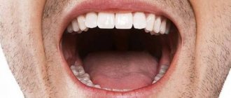
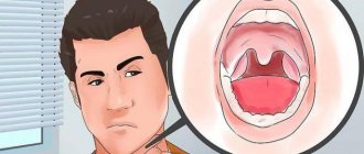
Visual examination should show enlargement and swelling of the salivary gland. It feels like it's inflamed. The patient complains of pain when chewing and swallowing. During palpation, the stone is sometimes felt; special sounding will confirm or deny the presence of a stone; X-ray - the image will show salivary stones even when palpation or probing showed nothing. He will show his location; sialography - injection of a contrast agent into the duct of the affected gland will reveal a stone; cytological examination of saliva - will tell about the absence or presence of inflammation in the gland.
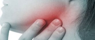
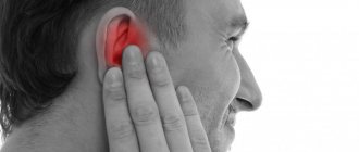
Symptoms of inflammation of the salivary glands
Regardless of which gland the inflammation occurs in, sialadenitis has the following symptoms:
- dry mouth associated with a decrease in the amount of saliva produced;
- shooting pain in the area of the affected gland, radiating to the ear, neck and mouth;
- pain when chewing and swallowing food, as well as when opening the mouth;
- swelling and redness of the skin in the projection of the inflamed salivary gland;
- unpleasant taste in the mouth, possible appearance of pus;
- in the area of inflammation you can feel a dense, painful formation;
- a feeling of fullness, pressure in the area of the affected gland may indicate the accumulation of pus in it; increase in body temperature to 39 C, weakness.
A particularly dangerous form of sialadenitis is mumps, often called mumps. The disease is dangerous because the virus that causes it can affect other glands of the body (mammary, reproductive, pancreas). In addition, mumps is a contagious disease, so if signs of inflammation of the salivary glands appear, the patient should limit contact with healthy people and consult a doctor to clarify the diagnosis.
If left untreated, purulent complications of the disease may develop. When an abscess of the salivary gland occurs, the patient’s body temperature rises sharply, and the general condition worsens. It is possible that the abscess may break through either into the oral cavity, or a fistula may form on the surface of the skin.
Treatment
Modern medicine has more than one method of getting rid of salivary stones.
- Conservative – it is possible in the first stages of the disease. This is drug treatment. Secretion stimulants and antispasmodics are prescribed. For severe pain - anesthetics, penicillin-novocaine blockade. According to indications - taking antibiotics. As an auxiliary agent, use ordinary dry heat. Physiotherapeutic procedures using the Sollux lamp with conservative treatment can alleviate symptoms. A special diet will also increase the amount of saliva produced.
- Operative - used if the conservative method did not help. It is also indicated in the presence of pus in the salivary gland. The surgeon makes an operational incision in the jaw at a certain angle above the affected duct and removes the stone and cleans the gland.
- Interventional sialendoscopy is an advanced method of treating this disease - salivary stones are removed using an endoscope.
- Extracorporeal lithotripsy is the most modern way to get rid of mineral formations - grinding them with ultrasound.
Treatment of salivary gland tumors
The main treatment method for malignant tumors of the salivary glands is surgery, since such tumors are little sensitive to radiation and chemotherapy.
If the tumor is small in size and has a superficial location, partial removal of the gland along with the tumor is performed. In patients with large tumors, the gland is completely removed.
Radiation therapy before surgery and in the postoperative period can be used for significant tumor sizes.
Chemotherapy with the drugs Adriamycin, Platidiam, bleomycin, and methotrexate can be prescribed for inoperable (unremovable) tumors, but the effectiveness of this method is minimal.