- Causes and risk factors
- Localization
- Metastases
- Symptoms of Oral Cancer
- Stages of the disease
- Treatment
- Prognosis and survival
- Prevention
Malignant tumors of the oral cavity account for about 6% of all cancers in general. Based on the histological structure (type of tumor-forming cells), the following types are distinguished:
- Tumor of epithelial cells - cancer
- Tumor of connective tissue cells - sarcoma
- Melanoma
Each type includes several varieties.
Several groups of precancerous diseases are also distinguished separately. Precancerous diseases of the oral cavity are divided into:
- Obligate - with a high incidence of malignancy. These include Bowen's disease, warty precancer, limited hyperkeratosis, Manganotti cheilitis.
- Optional - with a lower incidence of malignancy. This group includes the verrucous form of leukoplakia, papillomatosis, erosive-ulcerative and hyperkeratotic forms of systemic lupus erythematosus and lichen planus, post-radiation stomatitis and post-radiation cheilitis, keratoacanthoma.
Among malignant tumors of the oral cavity, cancer is the most common.
Oral cancer, in turn, is divided as follows.
- Intraepithelial carcinoma (Carcinoma in situ, cancer in situ) is characterized by the absence of invasion into the basement membrane, despite the malignancy of the neoplasm.
- Squamous cell carcinoma is the most common.
Anatomical structure
The oral cavity is the initial section of the digestive tract, in which food is chewed and saliva is produced to digest food. It is involved in the process of breathing, swallowing, articulation and speech.
The composition of the oral cavity includes:
- vestibule (lips, front side of teeth, inner surface of cheeks);
- gums;
- the bottom on which the tongue lies;
- two thirds of the tongue;
- teeth;
- retromolar triangle - the space on the lower jaw behind the third molar;
- hard and soft palate.
Prevention measures
Not all oral and oropharyngeal cancers can be prevented, but the risk of developing these cancers can be significantly reduced by avoiding certain risk factors.
Limit smoking and alcohol
Tobacco and alcohol are among the most important risk factors for the development of RPD. Not starting to smoke is the best way to limit your risk. But quitting tobacco also reduces the risk of developing cancer, even after years of use. The same applies to alcohol.
Preventing HPV infection
In recent years, vaccines have become available that reduce the risk of contracting certain types of HPV. These vaccines were originally used to reduce the risk of cervical cancer, but have been shown to reduce the risk of other HPV-related cancers such as penile, anal, vulvar, and vaginal cancers. HPV vaccination also appears to reduce the risk of RCC, although this has not yet been proven.
Limiting exposure to UV radiation
Ultraviolet radiation is a risk factor for the development of RPR. If possible, limit the time you spend outside during the middle of the day, when the sun's ultraviolet rays are strongest. If you are out in the sun, wear a wide-brimmed hat and use sunscreen and lip balm with a sun protection factor (SPF) of at least 30.
Maintaining a Healthy Diet
Poor diet has been linked to RCC, although it is not entirely clear which substances in healthy foods may be responsible for reducing the risk of these cancers.
Classification
Oral cancer is divided into three types:
- papillary. The nodule in the mucous membrane increases in size and hangs into the oral cavity. The neoplasm progresses slowly;
- infiltrative. The seal on the pinkish mucosa is distinguished by a whitish color, clear contours and shape, and thinning of the membrane around it. On palpation from the side of the cheek, a dense infiltrate is felt. The tumor tends to grow rapidly. The patient complains of unbearable pain;
- ulcerative The most common form of the disease. Ulcers on the mucous membrane do not heal, they grow, and the border around them turns red. The outline is torn and its edges are bleeding.
Tumor metastases appear quickly. Malignant cells grow into the mental, submandibular, and deep jugular lymph nodes. This process is influenced by the thickness and depth of the tumor. Thus, when the tumor deepens by 4-5 mm, metastases occur in 98% of cases. At the T1 stage of oncology, metastasis is detected in half of the cases, and when the T4 stage is reached, distant spread of cancer cells is observed in 85% of cases.
The mechanism of development of cheek cancer
Malignant tumors of the cheek occur against the background of precancerous changes in the epithelial and subepithelial layers (leukoplakia or erythroplakia). The risk of leukoplakia degenerating into invasive cheek cancer is about 4-6%, with erythroplakia it reaches 30%. Subsequently, dysplasia transforms into “cancer in situ”, which penetrates into surrounding tissues and metastasizes to local and regional lymph nodes.
In some patients, even those cells that do not initially raise suspicion of dysplasia upon initial microscopy may gradually become malignant. As the tumor process progresses, distant metastases occur in the bones, lungs, and liver. Buccal cancer can grow through the skin.
Causes
The prevalence of oral cancer is growing and is currently diagnosed in 2% of patients among the total number of cases. Since 2009, the incidence has increased by 25%, with mostly squamous cell carcinoma being detected and only in isolated cases adenocarcinoma.
Most foci of oncology are observed in the tongue. Slightly less malignant formations on the floor of the mouth. Cancer of the soft and hard palate, gums and cheeks is detected in 20% of cases. Much less frequently diagnosed is damage to the alveoli of the lower jaw - 4%, the arches of the palate, retromolar region and vestibule - 3%.
Based on practice, men are more susceptible to oral cancer than women. This is due to bad habits, for example, the abuse of cigarettes or chewing tonic mixtures increases the production of saliva, which washes away beneficial elements from the mucous membrane. The risk group includes patients with HPV, elderly people, workers in hazardous industries, patients with lichen planus, people whose oral mucosa is systematically injured by fillings, prostheses, and metal objects.
Treatment for cheek cancer
Malignant tumors are successfully cured using radical radiation therapy while preserving the function of the oral cavity. Radiologists implant radiation sources because it is possible to irradiate a small volume of tissue at a high dose. Radioactive isotopes of cesium, gold, radium, iridium, tantalum, which have the same efficiency, are used as sources.
For small tumors, the size of which does not exceed 1 cm, they are limited to implantation of a radiation source, without resorting to additional external influence. In cases of slightly larger tumors that are not suitable in size for implantation, external irradiation is used along with source implantation.
Traditionally, large cheek tumors are treated with external beam radiation. Recently, oncologists have been using a combination of radiation and chemotherapy. Mobile lymph nodes are radically excised. During prophylactic removal of lymph nodes without signs of damage, in a significant number of cases, foci of micrometastasis are found in them. For this reason, radiologists prefer to perform prophylactic irradiation of the neck in patients without evidence of lymph node involvement by the external beam, sometimes in combination with surgical treatment.
After surgery for cheek cancer, a cosmetic defect is formed. The results of surgical intervention are improved using the technique of microvascular free skin grafting. If a patient develops dry mouth after radiation therapy, he is prescribed oral pilocarpine. The drug increases salivation, which is usually accompanied by minor side effects - sweating and increased urination. For primary or secondary treatment of tumors of the buccal mucosa, patients are prescribed chemotherapy drugs.
Early diagnosis of a cheek tumor allows for effective treatment. If the tumor process is at a late stage, the prognosis worsens. If you experience unpleasant sensations in the oral cavity or identify ulcers of the buccal mucosa, undergo an examination at the clinic by calling the Yusupov Hospital.
Symptoms
A malignant ulcer from ordinary stomatitis in the mouth can be identified by swelling and swelling of the cheeks, pain and constant discomfort even at rest. You should be wary of prolonged non-healing of the wound and its bleeding.
As the disease progresses, the symptoms intensify:
- swelling increases and spreads to the neck;
- the red or white spot on the oral mucosa intensifies;
- discomfort when chewing and swallowing;
- difficulty speaking due to friction of the mucous membrane on the teeth when moving the jaw;
- the appearance of bad breath;
- feeling of a foreign object in the throat;
- anemia of the mouth.
In the late stages of cancer, teeth fall out and body weight rapidly decreases.
Causes and risk factors
The causes of the development of malignant neoplasms of the oral cavity can be divided into local and general risk factors.
Common factors include age, a history of various hazards (exposure to radiation, etc.), and hereditary predisposition.
Local factors are local factors affecting the oral cavity. These include chewing nasvay (tobacco- and drug-containing mixtures), smoking, the habit of drinking scalding hot drinks, chronic traumatization of the mucous membrane (tooth fragments, deformed dentures), as well as the presence of precancerous diseases. A separate risk factor for detecting the disease in later stages is the lack of annual dental examinations.
It is the neglect of preventive visits to the doctor that prevents the diagnosis of cancer at early, treatable stages, detection and treatment.
Diagnostics
At the initial consultation, the doctor examines the oral cavity, examines ulcers, erosions, damage to the mucous membrane, and then takes a smear for examination. To confirm the inflammatory process, the patient is sent for a general and biochemical blood test.
The diagnosis is confirmed by the results of the examination:
- MRI and ultrasound of soft tissues of the neck. The images reveal the localization of the pathology, the depth of germination and the structure of the tumor, compaction from blood and lymph, decomposition of the cortical layer of the bone;
- if metastases are suspected, a fine needle aspiration biopsy of the lymph nodes under the chin, under the jaw and in the upper third of the neck is performed;
- positron emission tomography. Shows the depth of the tumor, as well as early metastases;
- osteoscintigraphy. Skeletal bones are examined to look for displaced cancer cells;
- CT scan of facial bones with contrast. The images show the tumor growing into the neck vessels, jaw or base of the skull.
Symptoms of Oral Cancer
Manifestations of the disease depend on the nature of the tumor and the location. Typically, the affected area is either a local ulceration or an induration (bulge, nodule). The patient feels pain and discomfort in the corresponding area. Warning signs include the following:
- A long-term, non-healing mouth ulcer. The oral cavity is rich in blood vessels, the mucosal epithelium is well supplied with blood, and normally, in the event of injury, rapid healing occurs. Any damage to the oral mucosa heals faster than a similar injury to the skin of the arm or leg. Therefore, if there is an ulcer in the mouth and for unknown reasons it does not heal for a long time, you should seek medical help.
- Lumps, “balls” under the mucous membrane, regardless of the area of the mouth where they arose. A compaction under the mucosa can be a symptom of both a non-oncological pathology (for example, a cyst of one of the small glands) and a sign of cancer. Only a specialist can distinguish one from the other. The seal, which initially appeared as a cyst, may subsequently become malignant if left untreated.
- Impaired swallowing, chewing, discomfort at the root of the tongue - all this is a reason to suspect cancer in the corresponding area.
- Toothache and bleeding in a specific area of the gum may be signs of cancer. And, although similar symptoms are caused by much less formidable ailments, consultation with a dentist is necessary. Thus, any violation of the anatomical integrity or functionality of the organs of this zone carries a potential threat, and this is a reason to see a doctor.
Treatment
The choice of treatment tactics depends on the stage and extent of the tumor. When the tumor grows rapidly, treatment methods are combined.
Operation
The doctor determines the principle of surgical intervention after determining the stage of the tumor and its spread. If cancer cells have penetrated the periosteum and surrounding tissues, a wedge-shaped, planar or sagittal resection of the jaw is performed. If the examination reveals the growth of cancer cells directly into the bone or the defect is noticed during surgery, segmental resection of the lower jaw is performed. The doctor assesses the lesion on site and determines the thickness of the excised layer.
The next stage of the operation is partial or complete excision of the cervical lymph nodes to prevent metastases if the thickness of the tumor is more than 4 mm or the location of the tumor in the floor of the mouth or on the tongue. If the tumor is located in the midline, then the cervical lymph nodes are excised on both sides. The operation ends with the immediate replacement of damaged tissue.
After removal, the tumor is sent for histological examination. Its size, thickness, depth, edges are assessed. Further treatment is affected by cell growth beyond the boundaries of the capsule of the removed lymph node, and the spread of cancer cells to neighboring organs.
Radiation therapy
Radiation after surgery is prescribed when diagnosing T3, T4, N2, T3 stages of the disease no later than six weeks after tumor removal. The need for radiation therapy increases with perineural invasion of the lymphatic vessels. The total focal dose for all sessions is 60 g, and the single focal dose for one session is 2 g. When metastases are detected on the neck, the SOD increases to 66 g, and if there is no risk of metastasis, the SOD decreases to 50 g.
As the main treatment, radiation therapy is used in a total focal dose of 60-70 g. The procedure is performed five days a week and is combined with chemotherapy. Every three weeks, 100 mg of cisplatin is administered.
Chemotherapy
Anticancer drugs are prescribed before surgery or along with radiation therapy to reduce the size of the tumor. Sometimes therapy is prescribed simultaneously with surgery.
Treatment involves the use of a 5-fluoroacyl regimen together with cisplatin or other agents - carboplatin, methotrexate, bleomycin. They cause a number of side effects, for example, vomiting or nausea, hair loss, decreased appetite, and increased bleeding. Symptoms disappear after treatment, but permanent hearing loss is sometimes observed after taking cisplatin.
The prognosis of oral cancer depends on the stage at which the disease is detected. If treatment is started at stage zero, the disease will stop. It is worth noting that smoking provokes relapse or degeneration of the tumor, so repeated surgery or radiation may be required. Surgery at the first stage increases survival rate to 80-85%, and the combination of radiation therapy with surgery at the second stage by 60-80%. Already at subsequent stages of cancer development, the survival rate is no more than 50%, and all three treatment methods are used simultaneously.
Prognosis and survival
The prognosis of the disease depends on the stage, localization, and timely provision of assistance. In the early stages, with adequate treatment, the disease can almost always be overcome. When contacting a doctor was not timely, the prognosis is somewhat worse, and the percentage of cured patients is lower. In assessing the prognosis, the presence or absence of distant metastases is important. Despite enormous progress in oncology, distant metastases sharply reduce 5-year survival; this is a significant problem for a specialist. In addition, the prognosis depends on the histological type of the tumor, because The growth rate of different types of tumors can vary significantly. The age of the patient, the location of the tumor, the presence of concomitant diseases in the patient, the chosen treatment tactics - all these factors significantly influence the prognosis.
Dispensary observation
Since the tumor can recur and metastasize, after completing the course of treatment the patient is registered with the oncology clinic. The first year you should visit a doctor every month, the second year a preventive examination is carried out every 4-6 months, and then once a year or in case of any ailments. The examination involves an examination - ultrasound and contrast MRI of the soft tissues of the neck, PET, osteoscintigraphy. Consultation with an otolaryngologist, dentist and oncologist is required. The doctor may shorten the period of medical examination if there is a high risk of relapse.
List of references on the topic:
- Gantsev Sh.H. Oncology – M, 2012 – P.204-205.
- Golovin D.I. Errors and difficulties in diagnosing tumors, D.: Medicine. Leningr. department, 2015 305 pp.
- Selected lectures on clinical oncology/Ed. IN AND. Chissova, S.L. Daryalova. – M., 2010
- Matyakin E.G., Alferov V.S. Chemotherapy of head and neck tumors // Mat. 2nd Ros. oncol. conf. “Current trends in the development of drug therapy for tumors” December 8–10, 2021 – M., 256 p.
- Tumors of the head and neck: hands/ A.I. Paches. - 5th ed., add. And revised - M.: Practical Medicine, 2013. -478 p.
- Shine A.A. Oncology. M – 2014 365 pp.
- Encyclopedia of Clinical Oncology/Ed. M.I. Davydova. – M., 2014 –P.140-179.
- Bityutsky P.G., Kitsmanyuk Z.D., Trofimov E.I. Diagnosis and treatment of cancer of the oral mucosa // Medical consultations. - 2014. - No. 1. - P. 23-27.
- Byakhov M. Yu. Options for combined and complex treatment of locally advanced cancer of the oral mucosa and oropharynx: Dis. Dr. med. Sci. - M., 2013.
Stages of the disease
Stage I is characterized by the presence of a tumor up to 1-2 cm in diameter, not extending beyond the affected area (cheek, gums, palate, floor of the mouth), limited to the mucous membrane. Metastases are not detected in regional lymph nodes.
Stage II - a lesion of the same or larger diameter, which does not extend beyond any one part of the oral cavity, but extends into the submucosal layer. There are single metastases in regional lymph nodes.
Stage III - the tumor grows into the underlying tissues, but not deeper than the periosteum of the jaw, or has spread to adjacent parts of the oral cavity. In regional lymph nodes there are multiple metastases measuring up to 2 cm in diameter.
Stage IV - the lesion spreads to several parts of the oral cavity and deeply infiltrates the underlying tissues, in the regional lymph nodes there are immobile or disintegrating metastases, and the presence of distant metastases is also characteristic.
Classification by stages is periodically subject to revision; you can find a division of stages into subtypes - A and B. Currently, classification by stages is used less and less, the TNM classification is more relevant. Its principle is that this coding indicates the characteristics of the tumor itself, the condition of the closest (regional) lymph nodes, and the presence or absence of distant metastases.
In the diagnosis, the oncologist indicates the histological type of cancer, since different types of cells differ in growth rates, tendency to metastasize, and sensitivity to treatment. All types of classifications serve one purpose - to correctly assess the extent of the spread of the disease, the degree of damage and develop the right tactics of assistance.
Book a consultation 24 hours a day
+7+7+78
Tongue cancer
It is not the most common form among cancers, but still, it occurs quite regularly, accounting for the majority of cases among oral cancers.
Tongue cancer is a malignant tumor, the basis of which is the epithelial cells of the tongue mucosa. The disease is characterized by a diffuse or local type of compaction of organ tissue, papillomatous growths formed on the surface of the tongue, as well as ulcers.
The likelihood of tongue cancer occurring in different locations increases with age. Most people treated for this disease are men between 40 and 60 years old. After 80 years of age, this disease rarely begins to develop initially. There is evidence of tongue cancer being detected in young children, although this pathology is not typical for this age.
Causes and risk factors.
Among the reasons for the development of tongue cancer, the leading importance is given to external unfavorable factors:
- Long-term smoking and drinking alcoholic beverages. The combination of these two factors increases the likelihood of malignant neoplasms in the oral cavity several times: alcohol significantly enhances the carcinogenic properties of tobacco mixtures.
- Chronic mechanical injury to the tongue: wearing poorly installed dentures, when the mucous layer is injured by the edge of a broken tooth, or when regularly biting an organ.
- Occupational hazards - working with heavy metal salts and oil industry products.
- Constant consumption of dishes that are too hot, burning the mucous membrane of the tongue, constant use of hot spices.
- Chronic inflammation of the oral cavity - stomatitis, gingivitis, hyperkeratic forms of systemic lupus erythematosus, lichen planus.
! The simultaneous influence of two or three unfavorable factors on the human body increases the likelihood of cancer in the oral cavity.
There are also some precancerous diseases, with the development of which the risk of developing a malignant tumor of the tongue increases several times. These diseases include:
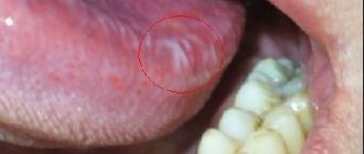
- Bowen's disease is the formation of a single spot on the tongue, its surface is smooth. The lesion may sink or erosion may form in its place (photo).
- Leukoplakia is an area of constant inflammation that may look like a whitish spot or a gradually growing wart. Such changes tend to become keratinizing.
Classification of tongue cancer.
Tongue cancer is divided according to its localization in the organ, shape, growth pattern and histological structure.
By localization (location):
- Cancer of the body of the tongue. This localization is detected in approximately 70% of patients; usually, with this location, the tumor affects the lateral surfaces of the organ or its middle part.
- Cancer of the root of the tongue is detected in 20% of cases. This localization is also referred to as oropharyngeal cancer. The occurrence of malignant tumors in the posterior half of the oral cavity always has a more aggressive course.
- A cancerous growth localized at the bottom of the tongue. Found in 10% of cases.
(tongue root cancer)
According to the form, tongue cancer is divided into:
- Ulcerative form. An ulcer forms on the tongue, with uneven and often bleeding edges. Ulcerations affect the lower part of the tongue and its middle.
- Infiltrative form. In the patient’s tongue, a lumpy, dense lump can be palpated – an infiltrate. The mucous layer above the seal is thinned, and with this form of cancer pain is pronounced. Infiltration in most cases forms at the tip of the tongue and in its back.
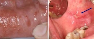
- The papillary form occurs when a tumor on a thin or thick stalk grows above the tongue in its different parts from the mucous layer. This type of malignancy is characterized by slow growth and usually affects the sides of the tongue.
(initial stage of papillary tongue cancer)
Depending on the growth pattern, tongue cancer can be:
- Exophytic tumor. In this case, a malignant neoplasm grows into the oral cavity.
- Endophytic tumor. The location of the formation is limited by the thickness of the organ.
Clinical manifestations.
In the development of tongue cancer, there are three stages of its formation:
- initial stage,
- developed
- launched.
Initial stage. It is characterized by an asymptomatic course, which may be invisible to the patient. The first stage is manifested by the formation of special papillary outgrowths or whitish spots on the tongue, which are often confused with ordinary plaque. In addition, local compactions and redness may form, which appear immediately on the side of the tongue. Enlargement of the submandibular lymph nodes may also occur.
As a rule, pain does not occur at the initial stage, but in rare cases it can be observed. If the pain is still characteristic of the initial stage, then it usually does not have a clear localization, therefore it is often perceived as caries, periodontitis, glossitis, pulpitis, traumatic glossalgia or a chronic type of tonsillitis.
Developed stage - expressed by various symptoms. It is often accompanied by characteristic pain of varying intensity. The pain can be of different localization and have a so-called diffuse nature. It happens that the pain syndrome can spread to other areas of the oral cavity or even to the ear.
When the oral mucosa is irritated by necrosis products, salivation may increase, and due to the disintegration of the tumor, patients experience bad breath. At this stage, difficulties arise when swallowing, the patient feels numbness of the tongue. In addition to pain when swallowing, there may be difficulties in speaking or pronouncing certain sounds.
Clinical manifestations according to the type of cancer:
- Papillary - presented in the form of a dense growth located above the surface of the mucosa, covered with plaque-like or papillary growths.
- Ulcerative - occurs less often among patients, almost 2 times less often than papillary. It is characterized by an ulcerative surface surrounded by a ridge, which increases in size from time to time. In the first stages of development, this form will not be characterized by pain. Only after a while, when the tumor reaches a certain size, pain and characteristic bleeding will be expressed. If the ulcer is infected or has additional inflammation attached to it, then the cancer may camouflage itself, making it difficult to diagnose.
- Infiltrative - will be characterized by the rapid growth of the tumor, directed into the thickness of the tongue, which is manifested by its compaction. This form is characterized by a diffuse nature. It can spread to the entire tongue, leading to poor tongue mobility. Deep slit-like ulcers will be characteristic of the infiltrative-ulcerative form of tongue cancer.
Advanced stage. The tumor is characterized by an aggressive course and very rapid growth, which is also accompanied by destruction of nearby tissues, as well as metastasis to the region of regional lymph nodes (mental, cervical, submandibular). Metastases at this stage of tongue cancer can be in the lungs, liver or brain.
General symptoms of an oncological process:
- feeling of weakness,
- feeling of chronic fatigue,
- loss of appetite,
- weight loss,
- the appearance of dysfunction of internal organs.
Diagnostics.
The asymptomatic course of tongue cancer at an early stage does not allow timely detection of the disease.
As a rule, a change in the mucous membrane at the initial stage, detected during a routine examination or during the treatment of caries, may not be perceived significantly, but may be mistaken for a common injury, but not for cancer. For the same reason, many cases of tongue cancer can only be diagnosed at the second or third stage.
Primary diagnosis, namely examination and palpation of the tumor, is carried out by an oncologist. An accurate diagnosis will be established only after obtaining and studying the data of a cytological examination of a smear taken from the surface of such an ulcer , as well as after a histological examination of the tumor through a biopsy.
In order to determine how much the tumor has grown inside the organ and/or other structures of the oral cavity (determining the extent of the oncological process), instrumental visualization methods are used: ultrasound of soft tissues, radiography, CT scan of the facial skull and orthopantomography (x-ray examination, which allows one to obtain a detailed image of all teeth with jaws, adjacent parts of the facial skeleton).
To diagnose the presence of metastases of tongue cancer, the following are used: lymph node biopsy, MRI and CT scan of the brain, chest x-ray, liver ultrasound or skeletal scintigraphy.
! Differential diagnosis of tongue cancer should be carried out with a benign tumor of the tongue, one of the forms of primary syphilis, tuberculous ulcer and leukoplaxia.
Treatment.
Combined - radiation therapy, surgery and chemotherapy.
Radiation therapy.
As a rule, it is carried out both before and after surgery. Radiation therapy is carried out in two ways: remotely or by interstitial irradiation. As for the latter, the primary focus and the area of metastasis are irradiated first. If the tumor is large, then this, like infiltrative growth, is considered an indication for a combined type of chemoradiotherapy.
Palliative radiation therapy is combined with chemotherapy among patients with distant metastases.
Surgical treatment.
It is carried out to eliminate the tumor. Surgical treatment will include tongue resection or glossectomy. If the cancer has already grown into the soft tissue down to the bottom of the oral cavity, capturing the bone structures, then the operation will be accompanied by resection of the affected tissue or jawbone.
Plastic surgery
In further periods of postoperative recovery, plastic surgery methods can be used to restore the lost structures of the maxillofacial area. If tongue cancer has metastasized to the lymph nodes, then they should also be removed.
Prevention.
- Preventive measures to prevent tongue cancer include:
- Conducting self-examinations of the mouth and tongue
- Undergoing preventive examinations by specialists
- giving up bad habits (not drinking alcohol and smoking)
- eliminating the causes that provoked injury to the mucous membrane of the tongue - good treatment of fillings after their installation, correct selection and installation of dentures, as well as treatment of dental chips
- Carrying out oral hygiene is also an important preventative measure.
Treatment methods in Medscan
To treat oral cancer, the Medscan Oncology Center uses advanced technologies. The radiation therapy department uses high-precision Varian True Beam equipment, which provides effective results with minimal damage to adjacent healthy tissues and organs.
For cancer of the oral cavity, chemotherapy appointments will be needed. Medscan implements international treatment standards, so our patients take only high-quality modern drugs.
Another advantage of the treatment process in Medscan is the availability of an outpatient mode of operation. Comfortable conditions allow you to quickly adapt treatment measures to your usual lifestyle.
Diagnostic methods in Medscan
The diagnosis of “oral cancer” in Medscan is made based on the conclusion of a council of specialists. An interdisciplinary approach to the problem eliminates errors in further therapy.
Oncopathology is diagnosed using the following methods:
- Inspection, palpation of the lesion.
- Carrying out a biopsy with histological (study of a tissue fragment of a tumor under a microscope) and cytological (assessment of the cellular elements of the tumor) studies.
- X-ray examination when the bones of the facial skull are involved in the oncological process.
To clarify the prevalence of metastases in neighboring organs and tissues, additional steps are included in the diagnostic plan:
- CT scan;
- ultrasonography;
- Magnetic resonance imaging.