04.11.2019
When a patient has a painful tooth removed, he feels relief. A few days after the operation, the wound heals, and all the troubles are left behind. But during the healing period of the injured gum, you should especially carefully monitor its condition. And if something white appears in the hole that formed after a person had a tooth removed, you should immediately contact your dentist and find out the cause of this phenomenon.
Natural causes of the appearance of white plaque on a wound formed after tooth extraction
After tooth extraction, an open wound remains in the oral cavity, through which, if a person did not have immunity, any pathogenic microorganisms would penetrate. But such protective reactions of the body exist, and they are triggered immediately after the root is removed from the hole.
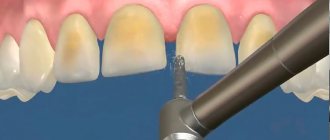
Human blood contains substances that, when the walls of blood vessels are damaged, form a fibrin clot that prevents bleeding. In the oral cavity, this process is also facilitated by special components of saliva. Part of the fibrin settles on the surface of the wound. In some people this layer is practically invisible, in others a visible white fibrinous plaque forms, like at the site of the pulled out tooth in the photo on the right.
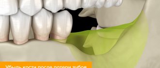
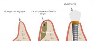
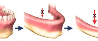
If tissue regeneration proceeds without complications, the fibrous plaque disappears over time. The void formed inside the hole is filled with epithelium and connective tissue, after which bone tissue is formed in this place. The healing process of bone tissue after tooth extraction is shown in the photo:
A few days after extraction, the gums near the socket become paler. This means that the inflammatory process, which always develops after surgery as a natural protective reaction of the body, subsides. The flow of blood to the wound decreases, and the color of the gum mucosa becomes the same.
Fibrin is an essential protein. Properties, functions, fibrin and inflammation
Contents
hide
1 Fibrin is an essential protein. Properties, functions, fibrin and inflammation
2 The role of fibrin in inflammation
3 Body protection
4 Function of fibrin
5 Experiments performed
6 Enzyme treatment
7 Method of treating patients with trophic ulcers
8 Classification of wounds. Wound process 8.1 Materials used in the article:
Fibrin is a protein that is the end result of blood clotting. It is formed as a result of the effect of thrombin on fibrogen.
Fibrin is an insoluble protein produced in the body in response to bleeding. This protein is a solid element consisting of fibrous threads. The progenitor of fibrin is fibrinogen. This is a protein produced by the liver. It is in the blood. When tissue is damaged, bleeding occurs. To stop it, thrombin begins to work. It affects fibrinogen, thereby provoking its transformation into fibrin.
First, the protein molecules combine into long strands that entangle the platelet, creating a rough mass. Gradually it hardens and contracts, forming a bloody clot. The compaction process is stabilized by a fibrin-stabilizing factor.
When white plaque at the site of an extracted tooth is dangerous
White plaque at the site of an extracted tooth does not always occur as a natural phenomenon. Sometimes this sign indicates a developing complication due to the dentist’s mistakes during tooth extraction or the patient’s improper behavior in the postoperative period:
- White spots near the socket, accompanied by redness and swelling of the gum mucosa, indicate infectious inflammation and suppuration, that is, the development of alveolitis. The socket becomes inflamed due to insufficient treatment with antiseptics during surgery, after injury or premature loss of a blood clot. If a sharp edge of the hole is formed, it will irritate the surface of the wound and open the way for microbes into the hard tissues of the jaw. Alveolitis occurs especially often after the removal of wisdom teeth, which is associated with increased traumatic surgery.
- If the tooth crumbles when grabbed with forceps, fragments of the root may remain inside the socket, as in the photo on the right. They prevent gum healing, injure surrounding soft tissues and can cause suppuration. Most often, fragments remain in the socket after wisdom teeth are removed, which is due to their uneven roots.
- If you chew food carelessly after tooth extraction, food particles can get into the socket and get stuck in it. After a few hours, they will begin to rot and become a source of infection.
Without appropriate treatment, inflammation of the socket can not only lead to the spread of infection to nearby tissues and internal organs, but also to the development of tumors.
Alarming symptoms
During the normal course of the healing period of the wound formed after tooth extraction, the unpleasant postoperative sensations pass quite quickly. The bleeding stops after about 3 hours (it may last longer if a wisdom tooth is removed), pain and swelling subside after 2-4 days, the blood clot and fibrous plaque resolve within a few days after extraction.
As complications develop, the following symptoms may appear:
- The pain intensifies, acquires a “shooting” character, and spreads to the jaw, ear or temple.
- Swelling increases, the lymph nodes in the facial area become swollen and hard.
- The bleeding does not stop after 3 hours, or bleeding resumes some time after the operation.
- A febrile state occurs.
- The sensitivity of the skin near the area of the jaw where the tooth was pulled out is lost.
- It becomes difficult to move your jaw.
- White or yellowish liquid flows from the hole.
- The wound is covered with a thick layer of plaque, which emits an unpleasant odor.
Enzyme treatment
Recently, special attention has been paid to the properties of enzymes. This is especially true for proteases. To treat fibrin enzymes, substances of this type are used. They help dissolve excess protein, thereby preventing serious complications such as blood clots.
The properties of proteolytic enzymes are different. They are able to have fibrinolytic and immunomodulatory effects on the body, as well as improve blood circulation and work as decongestants and anti-inflammatory substances.
Since thrombus formation is based on the production of fibrin, a protease is needed that causes the breakdown of this substance. Without such an enzyme, it is impossible to break the protein into molecules, therefore, there will be no improvement in blood microcirculation.
With local exposure to protease, it is possible to remove necrotic plaque, resolve fibrinous formation, and liquefy viscous secretions.
Is it necessary to remove white plaque formed on the gum after tooth extraction?
To decide whether it is necessary to take any therapeutic measures, you need to find out the reason for the appearance of white plaque on the postoperative wound by visiting the dentist again. If the doctor does not notice any signs of pathology, it means that the hole is covered with a natural fibrin coating. It cannot be removed because it is a protective barrier for pathogenic microorganisms.
If a person tries to remove fibrous plaque from the socket without consulting a dentist, he will provoke infection with further unpleasant consequences.
If the white plaque formed after tooth extraction is a sign of an inflammatory process, it is necessary to begin treatment as quickly as possible:
- If root fragments or stuck pieces of food are found, they are removed and the wound is thoroughly treated with antiseptics. The dentist may prescribe the patient a course of antibiotics so that the inflammation does not affect a larger part of the gum.
- If an infection develops, the doctor places a tampon with medicine in the hole. Oral medications may be prescribed, as well as antiseptic rinses.
- If a tumor has formed due to prolonged inflammation, it is removed surgically.
What not to do
If after tooth extraction a white plaque has formed in the socket, you should not begin self-medication before visiting the dentist. Attempts to provide first aid to yourself at home can lead to more serious complications:
| Patient Actions | Consequences |
| Mouth rinse | Washing off of the protective fibrin layer, loss of a blood clot with subsequent development of alveolitis |
| Trying to remove a clot, tooth debris, food or pus on your own | Infection that can spread not only to the soft, but also to the hard bone tissue of the jaw |
| Brushing the area of an avulsed tooth with a toothbrush | Damage to the fibrin layer, bleeding, infection of the postoperative wound |
| Applying a warm compress | Increased blood circulation leads to bleeding, and in the presence of suppuration, the formation of pus accelerates, increasing the likelihood of infection of internal organs |
| Taking medications | Individual intolerance reaction, unwanted side effects |
| Smoking in the first few hours after surgery | Burn of the injured mucous membrane of the gums, damage to the fibrinous layer and the development of the inflammatory process |
If the patient is able to contact his dentist, he can ask what pain and fever-reducing medications can be taken before visiting the dentist.
In most cases, you should not be alarmed by the white fibrinous plaque in the part of the gum where the teeth had to be pulled out. This phenomenon is a natural reaction by which the body protects itself from infections. But if the hole becomes covered with a thick coating with an unpleasant odor, swells and turns red, and the patient’s condition worsens, you need to visit the dentist again to treat any complications that have arisen.
Category: Tooth extraction Published by Mister stomatolog
The role of fibrin in inflammation
Fibrin production and inflammation are two closely related processes. Protein plays an important role in contact with deteriorating, damaged tissue. Thrombokinase released from the tissue comes into contact with fibrinogen.
When blood clots, all toxic substances become clogged in the clot. This feature of the effect of protein during the inflammatory process protects the body from the further spread of toxins and their negative effects. This reaction is called fixation. Thus, fibrin is also a protector of the body from toxins.
Method for treating patients with trophic ulcers
Owners of patent RU 2578382:
The invention relates to medicine and can be used to treat patients with trophic ulcers. To do this, the surface of the ulcer is treated with a swab moistened with saline solution. After this, cryotherapy is carried out with an applicator maximally cooled with liquid nitrogen to a temperature of -180°C with different diameters of the flat working surface. The exposure time is no more than 5 seconds. The method ensures bloodless, painless and rapid transformation of a chronic wound into an acute one and active closure of the defect, including by removing the necrotic component, fibrin films, biofilms from the surface of the ulcer, reducing colonization and contamination of microorganisms, removing phenotypically altered cells of the edge and base of the wound. 1 salary f-ly, 2 ill., 1 pr.
The invention relates to medicine, in particular to clinical and experimental medicine, and can be used in the treatment of patients with trophic ulcers and non-healing wounds.
Trophic ulcers, regardless of ethology, attract the close attention of various specialists. This is due to both the enormous material costs of treatment and the high social significance of this issue. Regardless of wound etiology, the principles of local management are largely similar. Adequately selected local treatment plays an important role. Currently, new high-tech dressing materials have appeared, the use of which, without a doubt, helps accelerate the healing of chronic wounds. However, the use of these agents without preliminary treatment of the wound is ineffective, since in all cases at the bottom of the ulcer there is fibrin, purulent-necrotic masses and single flaccid granulations in greater or lesser quantities (RU2388418, A61B 17/00, 2010; RU2340305, A61B 18/02 , 2008; RU2278689, A61K 38/17, 2006; RU2203061, A61K 31/47, 2003; Chernetskaya Yu.G. Application dosage form of hydrogels with miramistin and chymotrypsin. Man and Medicine, 2004, p. 848).
To clean the wound and prepare it for active closure of the defect by skin grafting or the use of collagen-containing preparations, the following treatment methods are used: surgical, autolytic, chemical and physical methods.
Classic surgical treatment should include removal of necrotic tissue, fibrin film, and excision of wound edges containing phenotypically altered cells. Surgical treatment transforms a chronic wound into an acute one, shortens the time to epithelialization, reduces exudation, and reduces the risk of infectious complications. It should be noted that full surgical treatment is impossible in the presence of soft necrosis and neuro-ischemic lesions on the extremities (ABI). The group of inventions relates to the pharmaceutical industry, namely to the use of pterostilbene (PTER), or pterostilbene in combination with quercetin (QUER), or any acceptable salt thereof for the prevention of skin cancer or for the prevention and/or treatment of skin diseases selected from psoriasis, atopic dermatitis and allergic dermatosis, by topical administration.