- Published by: Laima Jansons
Tumors of the ear and cheek can be different in structure and nature of origin. Some of them only visually spoil the appearance, without causing discomfort to the patient, while other pathological formations cause pain and deterioration of well-being.
It is impossible to cope with the problem on your own. A proper diagnosis will require a visit to the doctor's office.
Photo 1. A tumor in the ear area is a serious symptom that requires immediate medical attention. Source: Flickr (wellan)
Why do the ear and cheek underneath swell?
Swelling in the ear area can be caused by various diseases.
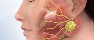
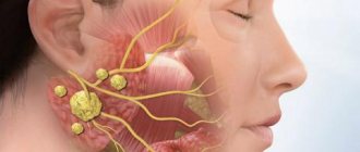
Inflammation of the lymph nodes
An important part of the immune system is the lymph nodes. They act as a biological filter that protects the body from the penetration of pathological agents (germs, viruses, fungi, bacteria). In case of infection with microorganisms the lymph nodes located behind the ears increase in size , which causes swelling that affects both the ear itself and the cheek.
This is interesting! Inflammation in the lymph nodes can also be caused by mechanical damage (trauma), neuritis of the auditory nerve, or the development of a tumor process in the body.
Atheroma
Compaction refers to tumor-like formations, divided into several types. Occurs as a result of blockage of the sebaceous glands , which is why a noticeable lump appears at the site of the lesion. Associated symptoms may include fever, redness around the formation, itching and stable, slow growth of swelling.
A capsule-shaped tumor is characterized by the presence of thick protein accumulations with an unpleasant odor. In rare cases, fluid leaks through the epithelium to the outside. The neoplasm is prone to degeneration into a malignant form.
It is important! Atheroma is detected in almost 8% of the population and older women are more susceptible to its formation.
Lipoma
A benign tumor, popularly called a wen . Pathology develops in connective muscle tissue. On palpation it is painless and easily movable. Lipoma is characterized by slow growth. The neoplasm is, rather, a cosmetic defect and does not pose a danger to life or health.
Mumps
An infection that penetrates the salivary glands causes large-scale swelling of the cheek in the ear area on one side. Mumps is characterized by an acute course of the disease, accompanied by an increase in temperature. Infection with the virus occurs through airborne droplets.
It is important! In childhood, the infectious disease mumps (mumps) is easily tolerated, while the lives of adult patients are in serious danger. Even death is possible.
Hemangioma
A benign formation consists of vascular tissue. The surface of the skin at the site of the tumor is lumpy and red or purple in color. The cause of compaction may be prolonged exposure to the sun, internal diseases or genetic predisposition.
In most cases, children and adolescents are susceptible to the appearance of hemangioma.
Other reasons
Skin abscess, rheumatoid nodules, lymphadenitis or mechanical trauma can also provoke the development of swelling in the ear area .
Causes
The etiology of ear cancer is studied by specialists from all over the world to this day. Scientists have put forward a theory that the oncological process develops under the influence of cancer viruses, which lead to DNA mutation. However, this assumption was only partially confirmed. Researchers have named the main factors that contribute to the formation of squamous cell ear cancer:
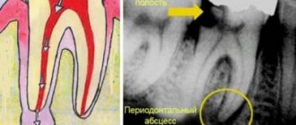
- the presence of chronic pathologies (otitis media, eczema);
- genetic predisposition;
- polyps;
- regular contact with aggressive carcinogens (alkalis, acids, heavy metals);
- inflammation of connective tissue.
It has been established that people who work in enterprises with radioactive radiation are most often exposed to the development of ear cancer.
Symptoms and signs of pathologies
Symptoms of the development of the pathological process differ depending on the nature of the origin of the tumor.
If there is an infection in the body , patients experience fever, weakness, pain when swallowing and palpating, as well as redness in the area of inflammation.
When infected with mumps, the patient develops pronounced swelling, and the face takes on a specific shape.
Oncological processes are painless. With a benign course, the person does not experience discomfort. In cases of cell degeneration into a malignant form, the tumor gradually increases in size, patients complain of decreased appetite and deterioration in general well-being.
Symptoms of jaw cancer
When a cancerous tumor originates in the upper jaw, there are practically no symptoms or they are identical to the manifestations of other types of pathologies, for example, sinusitis. That is why a malignant tumor can rarely be diagnosed in the initial stages.
With jaw cancer, patients experience:
- headache;
- numbness of the cheek;
- aching pain in the jaw;
- pungent odor from the mouth;
- purulent discharge from the nose.
Later, the following symptoms begin to appear:
- swelling of the neck;
- pain or numbness in the teeth closest to the tumor;
- loose teeth (a symptom of osteoporosis);
- alveolar processes may increase;
- a tumor from the bone of the upper jaw can grow into the orbit, causing displacement of the eyeball;
- neuralgic pain;
- headaches radiating to the forehead and temples;
- nosebleeds for no apparent reason;
- when the tumor reaches the trigeminal nerve, ear pain appears;
- mobility of the lower jaw is limited.
Sarcoma of the lower jaw is accompanied by the following specific symptoms:
- lower lip goes numb;
- contact teeth hurt;
- teeth become loose and fall out for no reason;
- bleeding ulcers appear on the mucous membrane;
- pain on palpation.
Homeopathic treatment
In the arsenal of homeopaths there is a whole range of drugs of natural origin, combining which can significantly improve the patient’s health and cope with the problem that has arisen. The specialist develops an individual treatment regimen based on the cause of tumor formation and accompanying symptoms.
The most popular drugs used to treat swelling in the ear area are presented below:
- Kali Bromatum . An anesthetic that stabilizes the flow of blood and lymph, and also reduces itching. Recommended for nervous and suspicious patients to normalize their general psychological state;
- Silicea . Normalizes the production of connective tissue and restores metabolic processes in it. Reduces swelling, eliminates suppuration and has analgesic properties. The product is effective for boils and carbuncles;
- Badjaga . Recommended for patients with weak nervous and cardiovascular systems to stabilize their general condition and eliminate benign tumors.
- Causticum . The most effective remedy for itchy nodules and swelling of cells under the skin in the ear area;
- Acidum fluoricum. Prescribed in the presence of benign formations, hemangiomas and tumors resulting from sunburn. Reduces itching, fever and improves immunity.
Photo 2. A homeopathic doctor will help you choose a medicine that suits your body.
Source: Flickr (Brenda McCutchen) The following multicomponent drugs also help get rid of a tumor on the cheek:
- Guna-Lympho is designed to accelerate the flow of lymph, reduce inflammatory processes in the lymphatic system and increase the protective functions of the body;
- Lymphomyosot has decongestant, immunomodulatory, detoxification and lymphatic drainage properties.
Treatment
The specificity of the disease is expressed in the fact that chemotherapy is not used for treatment - in this case it does not produce results. Gamma therapy is prescribed to stop the spread of the tumor and reduce its size. 3 weeks after irradiation, surgical removal of the jaw (resection or disarticulation) is performed. At a later stage, more extensive surgical intervention may be necessary: exenteration of the orbit, sanitation of the paranasal sinuses, lymphadenectomy.
Rehabilitation, restoration of speech, chewing and swallowing functions takes a long time. 2 years after complete recovery, correction is made using bone grafting methods, as well as using special splints.
Preventive measures
Since ear swelling is directly related to the immune system, it is necessary to maintain the immune system at a fairly high level, especially in the autumn and spring. To improve the body's protective functions, you should take vitamin complexes and normalize your diet by introducing more foods rich in useful minerals into your diet.
If you have the first symptoms of swelling of the cheek in the ear area, it is advisable not to delay going to the doctor . A small tumor may turn out to be a simple wen that does not require treatment, or a cancerous tumor that is life-threatening.
Try to avoid drafts and lubricate the wings of the nose with oxolinic ointment during mass epidemics.
Outer ear cancer
Ear cancer at the initial stage of development is asymptomatic. During the transition to stage 2, the sick person experiences severe itching and pain in the area of the ear cavity. Squamous cell carcinoma of the auricle is often accompanied by ringing or noise in the ears.
Upon examination, the doctor may detect a small ulcer or granulation in the area of the external auditory canal. Over time, the ulcers begin to bleed, burn and cause severe discomfort to the patient. In addition to bloody ulcers, the patient may detect serous and purulent discharge.
At stages 3 and 4, ear cancer metastasizes to neighboring lymph nodes. Cancer of the lymph node behind the ear is accompanied by severe pain, hearing loss, deformation of the facial nerves, and deterioration of the general condition.
Diagnostics
An accurate diagnosis is made after the following studies:
- otoscopy. A thorough examination of the ear cavity, eardrum, and ear canal. Otoscopy helps detect tumors in the external parts;
- histological examination. A diagnostic procedure to detect cancer cells in excised skin samples;
- MRI. One of the most accurate methods for determining the presence of cancer in the body. MRI allows you to determine ear cancer at stages 1 and 2 of development. In this case, the procedure allows you to find out more detailed information about the size, location and type of tumor;
- CT. An X-ray method for examining bone tissue for the presence of metastases.
In additional cases, audiometry may be required. This procedure helps to provide more accurate data on the state of the auditory analyzer.
Treatment
In the early stages of ear cancer development, the patient is prescribed courses of radiotherapy. After this, electroexcision is additionally performed. At stage 2, ear cancer is treated with surgical excision and subsequent radiotherapy. At later stages of development, radiation therapy with complete excision of the affected lymph nodes is performed to treat ear cancer. In case of multiple metastases, a Krail operation is performed.
Reasons for the development of atheroma
Atheroma develops in those places where there is an accumulation of a large number of sebaceous glands - in the area around the nose, on the eyelids, neck, scalp, chest, back and earlobe. Atheroma behind the ear is the most common of all types of atheroma, and, as a rule, affects both men and women equally.
The occurrence of atheroma of the earlobe is characterized by an unpredictable course and frequency of occurrence due to constant rubbing of the tumor with foreign objects - various hats, scarves, headphones, collars of shirts and blouses. In medical practice, cases of degeneration of a benign tumor into a malignant one are described, which occur in cases where proper surgical treatment is not carried out.
Symptoms of atheroma
Typically, atheroma reaches from 5 to 40 mm in size. In the initial stages, until infection sets in, atheroma may not bother patients. If suppuration occurs or the atheroma increases in size, the following symptoms may occur:
- Pain and redness in the area of atheroma.
- Swelling and increased body temperature.
- Burning and itching in the area where the atheroma is located.
- Feeling of free fluid when pressed.
In some cases, these symptoms of inflammation disappear within 10-15 days, after which the nature of the tumor changes. It becomes more dense, homogeneous and immobile. This occurs due to the replacement of the secretion of the sebaceous glands with connective tissue cells. At this stage, atheroma can give active growth, but can also remain in an unchanged position for several years. Metastasis to other organs is not observed in atheroma.
If a person’s immunity has enough strength to cope with the infection, then the atheroma spontaneously opens, as a result of which its contents drain from the cavity. This may be secretion from the sebaceous glands mixed with blood and pus. There is nothing wrong with this, except for the fact that after the autopsy, unsightly scars and scars remain on the body.
Of course, the presence of tumors such as atheroma of the earlobe or other area of the body should be an indication for surgical intervention and removal, since the tumor can grow into adjacent tissues and organs, as well as become inflamed as a result of a secondary infection.