Tooth extraction is a process that traumatizes the gums. After healing a diseased tooth, nerves, ligaments and small blood vessels are torn, which causes bleeding. This is a natural process - the blood does not allow pathogenic organisms to enter the circulatory system, washing them out. And then, curled up, it forms a barrier between unprotected tissues and bacteria. After tooth extraction, white plaque on the gum is not pus, but another stage of protection and restoration of the body.
When removing a tooth, the patient must be prepared:
➢
to moderate bleeding that may last for some time;
➢
the appearance of a dense blood clot in the gum;
➢
unpleasant odor 10-12 hours after surgery;
➢
the formation of a white coating on the blood clot;
➢
pain after surgery;
➢
temperature rise.
The listed symptoms are not a sign of an unsuccessful operation or infection in the body - this is the body’s natural response to tooth extraction.
What is white plaque on the wound after tooth extraction?
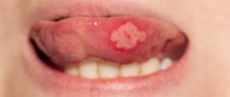
A few hours after the tooth is removed, a dark clot appears in the socket - bluish, black, red or brown. This clot is blood, and it is the primary protection of the socket from bacteria or pieces of food entering it. On the 2nd - 4th day, the patient may see that a coating appears on the clot - yellowish, gray or white. The deposit looks like pus and, together with the resulting bad breath, can alert the patient. However, there is no need to be alarmed - this plaque is not a sign of wound decay and indicates a normal healing process. This special protein compound is fibrin.
A milky plaque (fibrinous) can be very dense, hard or soft. This is no cause for concern. If you come to the dentist with this question for an unscheduled appointment, the specialist will tell you that everything is in order and there is no need to remove the film.
If the film is not removed and the wound is not disturbed, active processes will occur underneath it:
- the blood clot will begin to dissolve;
- the hole will begin to decrease in size;
- young cells, osteoblasts, will begin to move from the edge of the wound to the center;
- The gums will begin to close the wound.
Fibrinous plaque after tooth extraction is a natural stage of gradual recovery of the body, in which it is better not to interfere.
For what reasons does it appear?
White plaque in the mouth in adults occurs for the following reasons:
- development of endocrine pathologies;
- metabolic disorder;
- taking medications (antibiotics, hormonal drugs);
- vitamin deficiency;
- chronic infections of the nasopharynx and oral cavity;
- hepatitis (in particular hepatitis C).
Sometimes this symptom signals the development of cancer processes. In 10% of cases, intestinal cancer manifests itself this way.
What does it mean to have a whitish coating in your mouth in the morning?
In 80% of cases, this symptom is provoked by dry mucous membranes. This happens when a person sleeps with his mouth open - the appearance of deposits is due to a violation of natural hydration.
Another possible reason is impaired saliva production.
In women, this symptom occurs against the background of hormonal changes. Expectant mothers, as well as women in menopause, are at risk.
Appearance of yellowish deposits
A yellow coating appears in smokers, as well as in fans of black tea and coffee. If it is present in small quantities, there is no need to worry. If the deposits are poorly removed and are accompanied by additional symptoms, we may be talking about liver damage.
Possible reasons for the appearance of yellow deposits:
- gallstones;
- acute pancreatitis;
- cholecystitis;
- hepatitis.
A noticeable greenish tint may indicate stagnation of bile.
Why you don’t need to remove white plaque after tooth extraction
In some cases, patients intentionally or accidentally remove a blood clot or fibrinous film from the socket. This is not necessary, since removing a clot or film can provoke negative processes.
First of all, an open wound provides access for bacteria to the circulatory system and tissues of the maxillofacial apparatus. Infection can cause serious consequences and require long-term treatment with antibiotics. Also, removing the clot and white plaque increases the pain, since an open wound is more sensitive to any irritants, including drinking drinks and food, during a conversation.
Finally, plaque on the gums after tooth extraction does not need to be cleaned off because this can cause re-bleeding and prolong the wound healing stage.
In any case, there is no need to intentionally remove plaque; this can cause:
- serious complications;
- severe pain;
- improper gum formation;
- long recovery process after tooth extraction.
To avoid accidentally damaging the socket, blood clot or fibrinous film, you must follow the recommendations given by the doctor after the operation.
Establishing a diagnosis
If a plaque is detected on the palate of an adult’s mouth, the doctor undertakes:
- Listen to the patient's complaints.
- Collect anamnesis.
- Inspect the mucous membrane and skin.
- Make a microscopy of a scraping from the oral cavity.
Additionally, the patient may be referred for consultation:
- to a dermatologist - in order to identify fungal infections of other systems and organs;
- endocrinologist - to detect endocrine pathologies;
- an allergist - in order to determine the body’s sensitization to the materials from which dentures are made.
After clarification of the diagnosis, treatment is prescribed.
How not to damage white plaque after wisdom tooth removal
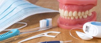
To eliminate the risk of complications and speed up the recovery process, you need to follow the rules of behavior after tooth extraction:
- do not eat or drink for 3 hours after surgery;
- stop smoking and drinking alcohol for a while;
- do not heat the cheek and gums, avoid overheating;
- for the first 24 hours, do not rinse your mouth or brush your teeth;
- Do not go to the bathhouse for a week, do not play sports.
Smoking is contraindicated, as it provokes vasospasm: the bleeding stops too quickly, the blood clot is not formed enough. Hot drinks, warming the cheek, sports training and visiting a bathhouse cause overheating of the body and dilation of blood vessels, which can cause re-bleeding, opening and infection of the wound.
Rinsing and brushing the mouth are acceptable 24 hours after tooth extraction, but they should not be overly vigorous. Hygienic procedures must be moderate so that the blood clot remains in the cavity and the subsequently formed fibrinous protective layer is not damaged. During the recovery period, food should be chewed only on the non-operated side of the jaw.
If you have a fever and severe pain in the first few days after tooth extraction, you can take Paracetamol - this will help reduce discomfort and improve your well-being.
Features of therapy
Treatment involves the use of topical drugs and internal medication.
To treat the oral cavity in adults, with very high sensitivity of the mucous membrane, it is allowed to use a diluted Lugol's solution (2 tsp per 200 ml of warm water from a kettle).
You can also lubricate the oral cavity with baking soda diluted in water (1/2 tsp per 150 ml of warm boiled water). The procedure should be carried out 2-3 times a day.
Drug therapy involves the use of antifungal and painkillers. Antihistamines are additionally used as prescribed by an allergist.
Use of antifungal agents
The main antifungal drugs are presented in the table:
| A drug | Description | Mode of application | How to use |
| Ketocanazole, 200 mg | The main effects are antiandrogenic, antifungal, fungicidal, fungistatic. The drug helps inhibit the synthesis of ergosterol, triglycerides and phospholipids. Against this background, fungi lose their ability to form colonies and threads. | Oral | The daily norm is 1-2 tablets for 14 days. Then, after the disappearance of acute symptoms - 1 tablet 24 hours - until the white deposits and ulcers completely disappear. |
| Fluconazole, 50 mg | The main effect is antifungal. Has a highly specific effect. | Oral | 50-100 mg/24 hours, for 1-2 weeks, until the sensation of plaque in the mouth completely disappears. |
| Nystatin, 1% | Effects – antifungal, fungistatic. The presence of double bonds in the composition helps ensure high tropism of the antibacterial drug to the sterols of the fungal cell membrane. | Local impact | The oral cavity should be treated 2 times/24 hours until the white plaque and other symptoms disappear. The approximate duration of the course is 10 days. |
| Clotrimazole, 1% | The product belongs to the group of imidazole derivatives. Effects – antibacterial, antiprotozoal, trichomonacid, broad-spectrum antifungal. | Local impact | The oral cavity is treated 2 times/24 hours until the white plaque disappears. The average course duration is 7 days. |
| Chlorhexidine bigluconate, 0.05% | The main effect is antiseptic. | Local impact | Intended for the treatment of mouth ulcers. 3 times/24 hours, 7 days. |
When white plaque on a wound after tooth extraction requires examination by a doctor
Despite the fact that white plaque after wisdom tooth removal is a natural reaction, the patient needs to conduct a daily self-examination of the oral cavity and consult a doctor for help if alarming symptoms appear.
You should consult a doctor if:
➢
on the 3rd - 4th day the pain does not decrease, it becomes throbbing and intense;
➢
a white, reddish or yellow mass or liquid is released from the wound;
➢
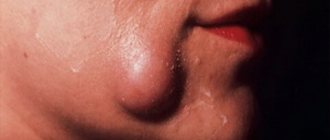
swelling of the gums has intensified or spread to the face;
➢
body temperature rose above 38 degrees.
These signs indicate possible infection or complications. After examining the oral cavity, the dentist, if necessary, may prescribe a course of antibiotics or surgical cleaning of the socket cavity.
If the oral cavity is covered with a white coating
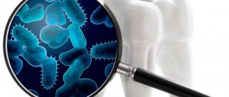
Teeth, probably more than any other structure in our body, can cause a lot of trouble if not taken care of properly. This happens because they carry the beginnings of many diseases of the digestive system, and can also significantly reduce immunity, especially if they are constantly exposed to all kinds of inflammation. The importance of timely prevention of dental diseases is no longer disputed by anyone. The oral cavity is especially susceptible to many diseases. Among them, there are diseases with fairly similar symptoms, the main of which is the appearance of white plaque on the mucous membrane of the gums, cheeks and tongue.
We list the diseases associated with the appearance of white plaque in the oral cavity :
- pachydermia of the mouth; - leukoplakia; - white spongy nevus of Cannon; - Lichen planus; — candidiasis (thrush); - chemical burns.
Candiosis and leukoplakia, just like oral pachyderma and chemical burns, are localized in any part of the oral cavity. Lichen planus can also occur anywhere in the mouth, but most often appears on the surface of the mucous membrane of the cheeks. White spongy nevus of Cannon is localized only on the surface of the buccal mucosa.
Let's talk in more detail about the features of these diseases.
Thus, oral pachyderma is usually caused by the presence of a persistent irritant. The raised, white lesion is caused by hyperkeratosis, a thickening of the oral epithelium secondary to healthy mucosa. When the irritant is removed, healing occurs, lasting for two to three weeks.
If a white spot is found in the oral cavity, and sometimes a convex plaque with clearly defined edges, these are characteristic signs of leukoplakia. When this disease manifests itself in a more severe form, the designated area will become compacted and bumpy, and cracks and erosion may appear on it over time.
Smoking is most often to blame for the occurrence of leukoplakia. So smokers need to be more careful about caring for and maintaining order in their oral cavity. In addition, leukoplakia can appear due to thermal, chemical, and mechanical irritants. Leukoplakia is often a precancerous disease, since the most severe type of this lesion can develop cancer. Fortunately, diseases are almost always treatable. At the same time, the plaque that occurs during this disease, unlike the same candidiasis, is not removed by simple scraping.
Candidiasis, or thrush, is a greasy white patch that occurs in sick children, frail elderly people, and in patients receiving high doses of corticosteroids or broad-spectrum antibiotics, or those suffering from acquired immunodeficiency syndrome (AIDS). Candiosis responds well to antifungal therapy after eliminating predisposing factors.
Cannon's white spongy nevus most often affects the cheeks symmetrically and can be treated without much difficulty, as does lichen planus, the grayish-white papules of which can even disappear spontaneously.
Remember that only a firm hand and the experience of a specialist accumulated over time will effectively save you from such diseases. If you have similar symptoms or suspicions, do not try to self-medicate - contact your dentist. The Apollonia dental clinic, located next to the Chertanovskaya metro station, offers a wide range of dental services in the treatment of diseases of the teeth and gums, as well as providing services such as prosthetics, implantation, teeth whitening and much more. You can consult with specialists by calling the numbers listed in the “Contacts” section.
Gum healing
After tooth extraction, you need to be prepared for the fact that pain and swelling may intensify on the second day after surgery. But already on the 3rd - 4th day after the operation, the discomfort will begin to decrease. By this time, the wound healing processes are more active, and by touching the tip of the tongue to the hole, you can feel that a compaction has formed.
During this period, the following are actively developing:
- bone formation in the area of the removed tooth root;
- narrowing of the socket due to the “growth” of the gums;
- formation of the mucous membrane in the socket area.
Fibrinous plaque after tooth extraction persists for a week. It disappears on its own, without mechanical cleaning. Little by little, the mucous membrane begins to turn pink, gradually acquiring a healthy, natural color. Around the 10th day, wound healing is completed: healthy tissue is formed that covers the mouth of the hole. A small depression remains in the area of the removed tooth. The formation of bone tissue at the site of tooth extraction takes much longer - up to 6 months. Changes will be visible on an x-ray.
Use of painkillers
The table shows the most effective analgesics. They are prescribed only according to indications.
| A drug | Description | Mode of application | How to use |
| Lidocaine hydrochloride, 1% | A potent local anesthetic. Effects – antiarrhythmic, local anesthetic. | Local impact | Prescribed against the background of hypersensitivity to amide anesthetics. Used for applications, before meals. |
| Procaine, 5 mg/ml, 0.5% | Local anesthetic. | Local impact | In the form of applications before meals and treating the mouth with antifungal drugs. |