What does anastomosis with the maxillary sinus mean?
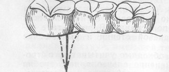
In the practice of surgeons, a complication often occurs that arises directly as a result of the removal of teeth in the upper jaw - the presence of a hole in the bottom of the maxillary sinus. The emerging communication between the oral cavity and the maxillary sinus requires the doctor to take urgent action to close the defect, since the oroantral communication can serve as a door for infection. The anatomical structure of the paired paranasal sinus is such that it is located close to the upper teeth and occupies almost the entire space of the upper jaw bone.
Treatment of the maxillary sinus consists of closing it and clearing it of mucus and other bodies. This operation is called sinusrothymia.
Application of mucoperiosteal flap
This technique is specially designed to close the pathology of the oro-ontral communication of the sinuses with the oral cavity.
The procedure requires the experience of a specialist, accuracy and strict adherence to medical protocol during the operation.
Main differences of the method
The main differences of the technique lie in the principle of its implementation, during which the surgeon cuts out a periosteal fragment of a trapezoidal mucous tissue flap.
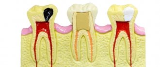
If there are appropriate indications, a maxillary sinusotomy is performed. Tissues that have undergone pathological changes and foreign elements are removed from the internal contents of the sinuses.
The edges of the communication are refreshed, curettage is performed and the hole is treated with antiseptic compounds.
A characteristic feature of this option for eliminating the anomaly is the two-layer delimitation of the message:
- first they form a lining from the inside of the sinus , placing a membrane on the mouth;
- then the next layer is formed by partially displacing the flap into the defect area.
After this, everything is sutured according to the U-shaped principle, which significantly accelerates tissue healing.
Expected Result
As a result of surgical intervention, it is possible to achieve:
- formation of higher quality layers;
- reduce the possible risks of repeated recurrent manifestations several times;
- completely eliminate the need to repeat such operations.
The method is indicated for the treatment of messages against the background of perforated sinusitis of the upper jaw, since it has the highest result in comparison with analogue methods of treatment practiced in domestic dental clinics.
The video shows the progress of surgical treatment of the oroantral communication.
When is anastomosis closure required?
Damage to the integrity of the voids of the appendages, which are located on both sides of the nose, can occur during the removal of a tooth from the alveolus in the upper jaw.
The channel occurs due to the following reasons:
- quick removal of a dental unit using surgical forceps;
- an operation to implant an implant into the jaw bone tissue;
- urgent treatment of a complex tooth with inflammation;
- chronic periodontitis, in which the ligaments and plate of bone that hold the tooth in the socket become thinner and flake off.
When performing manipulations in the upper row of the jaw, a cavity or fistula may form. The danger is the possible spread of purulent fluid. The problem is solved by surgery.
Clinical picture
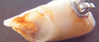
The oroantral communication is the connection between the maxillary sinuses of the nasopharynx and the oral cavity. In the hole formed as a result of the amputation of the tooth, a foamy liquid with bloody impurities begins to form.
In addition, there is pronounced bleeding of varying degrees of intensity from the nasal passage corresponding to the location of the removed dental unit. At the same time, the patient complains that he cannot whistle and puff out his cheeks.
The taken naso-oral sample has a positive result.
Diagnosis of perforation of the maxillary sinus floor
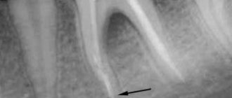
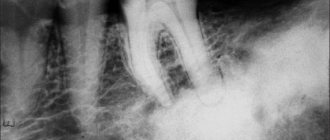
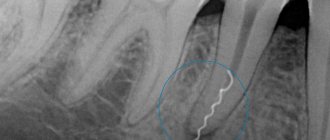
Checking for perforation of the maxillary sinus floor involves a comprehensive analysis of the typical clinical picture. It is necessary to take into account the condition of the socket of the extracted tooth, the presence of air bubbles, and bleeding from the nasal cavity. An X-ray examination is done, which shows the area of the hole with high accuracy.
The dentist may prescribe a procedure to insert a probe into the perforated canal. This will determine the presence of a bone bottom in the wound. The probe should pass through soft tissues without hindrance, without stopping or clinging to foreign bodies.
X-ray analysis will allow you to see darkening, characteristic of the presence of an accumulation of blood clots. In addition, X-ray images clearly show filling materials, implants, and fragments of tooth roots. Often, radiography uses a bright substance to clearly see the condition of bone and other types of tissue. To do this, the substance is placed into the cavity through the fistula.
An instrumental diagnostic method – computed tomography – will help determine the degree of perforation and the presence of foreign bodies.
If perforation is suspected, general clinical tests are prescribed to help determine the existence of infection in the human body.
Prevention
What needs to be done to avoid this problem? According to practicing dentists, the main thing a person can do to help themselves is regular visits to a specialist and timely sanitation of the oral cavity.
This simple measure will prevent the development of periodontitis and preserve the structural content and integrity of the bone septum.
In addition, after amputation of a tooth, it is advisable not to scrape the bottom of the socket to remove focal fragments.
When performing perforation, the surgeon should bring the edges of the gum as close as possible over the socket. This creates a favorable environment for the formation of a blood clot inside the cavity. It is this that will serve as a reliable barrier to the penetration of infection and pathogenic microorganisms that provoke inflammation.
What else can you advise patients? Take your own health more seriously and trust it only to professionals.
It is necessary to carefully choose a clinic, since one of the most likely reasons for this phenomenon is the unprofessionalism of the doctor.
How to treat perforation of the maxillary sinus floor?
A decisive preventive action that will prevent the inflammatory process of the maxillary sinus when an anastomosis has formed is the immediate closure of the communication. Closing the anastomosis with the maxillary sinus is performed using a trapezoid-shaped mucoperiosteal section formed at the beginning of the oral cavity, so that it is possible to peel off the flap a few millimeters above the transitional fold.
Before laying the mucoperiosteal area, the sharp edges of the alveolar process are smoothed with a special tool, parts of the flap and soft tissues from the side of the palate are cut to a width of 1-2 mm for better connection and better conditions for healing by primary intention. Before starting the procedure, the wound is treated with penicillin or streptomycin.
To ensure the safety of the flap, it is necessary to cover it and the adjacent tissue with gauze and a protective plate.
If a maxillary sinus anastomosis occurs during removal of the apical part of the root, removal of wisdom teeth, or during surgery for jaw cysts, it is necessary to suture the wound.
In case of perforation of the maxillary sinus during resection of the root apex, removal of wisdom teeth, or during operations on jaw cysts, the wound must be closed using closely spaced sutures.
Treatment options
To eliminate the pathology, several surgical options are used. The specialist will decide which one to prefer based on the clinical picture, the cause of the communication lesion and the anatomical features of the structure of the maxillofacial apparatus.
Relocation of the mucoperiosteal flap from the cheek
The main objective of this manipulation is to simplify the procedure for eliminating the message as much as possible and reduce the degree of injury at the time of surgery, while ensuring a high efficiency rate from the technique.
The doctor’s algorithm of actions is similar to the formation of a trapezoidal periosteum flap and is also carried out in two stages. The stages of the operation are discussed in more detail below.
The disadvantage of this method is the need to collect graft material and then attach it with sutures in the communication area, which is classified as an additional injury.
Use of membranes
Under local anesthesia, a periosteal flap fragment is initially cut out and slightly shifted upward. The frontal wall of the sinus is opened slightly and trepanation is performed. Then a slight maxillary sinusotomy is performed and foreign elements are amputated.
The area is well tamponed, the turunda is placed so that its end protrudes outward from the nasal passage.
In order for the flap to have mobility, the surface is slightly incised at the base. The wound is treated with antiseptics, and the mouth of the hole is covered with a special membrane.
The device is carefully inserted under the gum in the affected area from the palate so that it completely covers the defect by at least 3 mm, after which the device is pressed tightly for several minutes.
The working area is sutured with an interrupted suture. The method is effective and gives good positive results. Its advantage is the ability of the membrane to self-resorb.
Indications and contraindications for surgical tooth reposition, details of the operation.
In this publication we will talk about the features of mandibular sequestrectomy.
Here https://www.vash-dentist.ru/hirurgiya/operatsii-na-zubah/sut-osteosinteza-chelyusti.html detailed information about osteosynthesis of the lower jaw.
Plastic surgery of the anterior wall of the frontal sinus
The essence of the method is that in the process of anesthetizing the cavity, a submucosal resection of the nasal septum is first performed.
After this, under endoscopic observation, a luxation of the middle concha and a retrograde incision of the lower element of the uncinate branch are made, opening access to the frontonasal anastomosis.
This zone is the site of localization of polyps - during the process of plastic surgery of the walls of the frontal sinus, they are carefully removed along with thickened layers of mucous cells.
After this manipulation, a cavity arcuate incision is made in the direction from the middle of the eyebrow to the transition to the nasal slope and the periosteum is carefully dissected. The moved away soft fragments will expose the frontal sinus.
After the formation of a bone defect in this area, a revision of the sinus is made with a special medical chisel, which is quite sharp. Next, the affected mucous membrane, granulant, polyps, and purulent accumulations are removed, while trying to preserve as many healthy areas of soft areas as possible.
Finally, they ensure sufficient patency of the nasal canal at the mouth using a special catheter fixed in the sinus. Everything is done under the control of an endoscope.
If everything is done correctly and no complications have arisen, the tissues of the periosteum and mucous membrane are sutured, and a cosmetic suture is applied to the surface of the face to preserve external aesthetics.
Preventive measures before and after sinus lift to prevent the development of a fistula
Fistula after sinus lifting and bone grafting is quite easy to prevent by taking certain groups of medications before and after surgery, as well as observing the rules of oral hygiene.
Rules of conduct after surgery
The protective regimen should last from 5 to 7 days after the sinus lift. It includes a number of behavioral features:
- When brushing your teeth, special dental pastes with antibacterial and anti-inflammatory properties are used: Solcoseryl, Lakalut, Sensodyne. Oral hygiene procedures should be carried out twice a day (in the morning and before bedtime). It is necessary to brush your teeth on the operated side with great care so as not to disturb the condition of the tissues and implant after surgery.
- When eating, chewing movements should be carried out exclusively on the healthy side. Drinking all kinds of liquids through a straw is strictly contraindicated. You should also give up sweet foods for a week.
- During the first two weeks, you must stop playing any sports. This is especially true for athletics, gymnastics, cycling, swimming and martial arts. You should avoid the latter for a month.
- Try, if possible, not to sneeze or cough, avoid head injuries and bruises, and shaking.
- When sleeping, do not lie on your operated side.
Expected Result
If the removal of the formed canal between the oral cavity and the sinuses is carried out correctly, the patient will:
- inflammatory processes in the maxillary sinuses will decrease;
- purulent accumulations caused by parts of food and drinks entering the oral cavity will go away;
- be able to breathe freely.
The solution to eliminating unpleasant odors also applies. Sensitivity will return to the area of the former meatus and upper lip.
Reviews
Regardless of what caused the formation of the pathology, as well as the developed degree of zonal damage to the oral cavity and nasopharynx, the capabilities of modern medicine make it possible to qualitatively solve the problem, minimizing possible complications.
If you are interested in the method of treating oro-ontral communication, you can leave your feedback in the “comments” section.
If you find an error, please select a piece of text and press Ctrl+Enter.
Tags: operations in dentistry
Did you like the article? stay tuned
Previous article
If you have alveolitis, Neoconus in dentistry will cure it instantly
Next article
What is retrograde filling and features of its implementation
Symptoms of the disease
Some of the first manifestations of a fistula may be pain in the operated area when tilting and turning the head, a feeling of loosening of the implant, and an increase in temperature to 37.0 - 37.5 ° C.
The rate of progression of the disease is individual; over time, the denture may fall out (during its installation) or a hole may directly appear in the alveolar process. At this stage, there may be discharge of pus and mucus from the fistula, and a sensation of air vibrations in the oral cavity during breathing. When microorganisms or pieces of food enter the maxillary sinus, sinusitis develops, which aggravates the patient’s general condition. There may be a feeling of “softness” of the palate and alveolar process. If the fistula tract is 1 - 3 mm in diameter, then all kinds of discharge may be absent.
Eating becomes difficult due to severe pain. When coughing or sneezing, mucus may be thrown into the oral cavity, and there may be a sensation of air moving from the fistula.
One of the common signs of the oroantral tract is the development of odontogenic sinusitis. This disease can lead the patient to an otolaryngologist, directing the diagnostic algorithm and treatment in the wrong direction. This type of sinusitis is characterized by a long course with no response to treatment and antibiotics used. Puncture of the maxillary cave will also not have the desired effect.