Constant nasal congestion, persistent runny nose... We all know how unpleasant and debilitating such a condition can be. Meanwhile, the reason does not always lie in the runny nose or cold itself. One of the factors that provoke constant nasal congestion may be hypertrophy of the nasal turbinates. Symptoms of turbinate hypertrophy are very similar to those of allergic and chronic rhinitis. Treatment of turbinate hypertrophy depends on the cause of the development of this condition, and may include both conservative and surgical methods. We will talk in more detail about the nature, symptoms, as well as methods of treatment and diagnosis of this disease below.
The turbinates are spongy bony structures located in the nasal cavity on the side wall that guide and regulate air flow in the nasal passages. A person has three such paired bony outgrowths on both sides of the nasal septum, which are divided into upper, lower and middle. The turbinates are covered with ciliated epithelium, a special type of mucous membrane that cleans and moisturizes the air passing through the nose. The epithelium also serves as an immunological defense against bacterial and chemical irritants, its cells causing a rapid immune response in the form of an inflammatory reaction to the first signs of microbial or chemical irritation.
Why does turbinate hypertrophy occur?
The mucous membranes of each turbinate contain a large number of blood vessels and this means that they can easily shrink or swell in response to various factors. As a result, dilation of the turbinates can occur due to an allergic reaction, chemical or physical irritants, temperature changes and infections that enter the nose with air. This expansion is reversible, and usually the turbinates quickly return to normal size, but sometimes it can become chronic, which is called turbinate hypertrophy.
Headache - cyst is to blame
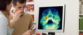
Small cysts in the maxillary sinuses do not manifest themselves in any way. The patient can only find out about them by chance, for example, during an examination for sinusitis.
Another thing is grown-up education. Reaching the size of the nasal sinus, the cyst rests against its walls and compresses the nerve endings. The patient begins to complain of facial pain radiating to the temples, eyes and teeth. Often, not understanding what’s wrong, a person first turns to a dentist or ophthalmologist.
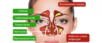
Further more. Extending beyond the maxillary sinus, the cyst compresses the trigeminal nerve, which causes severe headaches. Swelling appears on the face. Due to the displacement of the nasal wall, breathing on the affected side becomes difficult. In the worst case, it can result in otitis media, bronchitis and meningitis.
SYMPTOMS OF NASAL TUNCHA HYPERTROPHY
Characteristic symptoms of turbinate hypertrophy include:
- Difficulty breathing through one or both nostrils
- Chronic sinus infection
- Snore
- Frequent nosebleeds
Forewarned is forearmed
See also Treatment of ENT diseases Removal of a cyst in the nose Treatment of a maxillary sinus cyst Surgery to remove a cyst in the maxillary sinus
Following simple preventive measures will help to avoid the appearance of cysts in the right or left maxillary sinus:
- Monitor your immune system and try not to catch a cold. In frosty weather, refrain from visiting the pool and playing outdoor sports.
- If you get sick with ARVI, don’t let the disease take its course. It is viral diseases that become the main cause of chronic sinusitis, and after them - cysts.
- Visit your dentist regularly. Sometimes inflammation from the roots of the teeth spreads to the sinuses.
- After treatment, cysts may appear again. To prevent relapse, see an otolaryngologist regularly. Find out what is causing the problem. You may need to correct a deviated nasal septum or get rid of polyps.
And remember, any pathology must be treated by a specialist. Home treatment is ineffective and can have serious consequences.
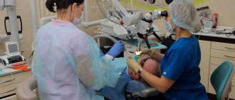
Radiofrequency turbinate reduction (RTR)
During this procedure, the doctor performs local anesthesia of the nasal cavity, after which the patient is positioned on the treatment table and the doctor performs radiofrequency reduction of the nasal turbinates using a special electrode. The principle of operation of RTR is based on short-term heating of the tissues of the nasal concha with an electrode, which leads to their compression and expansion of the respiratory passages.
What a patient should expect when undergoing the procedure in Singapore:
- Preparing for RTR only takes 15-20 minutes;
- The procedure is mostly painless and minimally uncomfortable;
- Immediately after the procedure, the patient may not experience significant relief from congestion due to slight swelling and crusting. However, immediately after the cleaning (which is performed by the doctor a few days after the procedure), the patient will be able to experience the full benefits of radiofrequency turbinate reduction;
- The procedure does not require hospitalization - the doctor performs it on an outpatient basis in the clinic. Immediately upon completion, the patient can return to his daily activities;
- The period of complete recovery and healing takes about 4-6 weeks;
- During the first few days after the procedure, patients are not recommended to swim in the pool.
Treatment
This disease has two fundamental approaches to treatment: conservative (non-surgical) and surgical treatment, i.e. removal of adenoids. Conservative treatment involves the use of desensitizing medications, as well as the local use of antiseptic drugs in the form of drops or sprays. Oily and iodine-based solutions can be used. The use of homeopathic treatment is also highly effective, which, like other drugs, is selected individually, based on the duration of the disease, the frequency of relapses and the age of the child.
Friends! Timely and correct treatment will ensure you a speedy recovery!
In addition to drug therapy, it is highly effective to conduct courses of physiotherapeutic treatment, consisting of laser therapy, ultraviolet irradiation (UVR), vibroacoustic therapy sessions, etc. All of the above methods are carried out absolutely painlessly and do not cause psychological trauma to the child. The number of treatment sessions can vary from 5 to 15, but for the physiotherapeutic effect to occur, at least 5 sessions must be performed.
If there is no effect from the ongoing drug therapy and physiotherapeutic treatment, and the child’s degree of adenoid vegetations is at the level of III degree or even higher, it is worth considering performing an adenotomy - partial removal of adenoid vegetations, or adenectomy - complete removal of the adenoids. Currently, operations using endoscopic technology are effective, which is very significant for an ENT doctor, since an ENT surgeon has the opportunity to introoperatively (during the operation) see how well this ENT operation was performed, whether the tissue of the pharyngeal tonsil was completely removed and the bleeding was stopped.
To minimize stress on the child’s part, it is recommended, if possible, to perform this operation under short-term anesthesia.
Make an appointment right now!
Call us by phone or use the feedback form
Sign up
Control of the effectiveness of the adenotomy operation is the complete relief of all complaints and symptoms after the operation, as well as the absence of adenoid vegetations during a control endoscopic examination.
Surgical treatment of turbinate hypertrophy
Surgery is used if drug treatment is ineffective. The purpose of the operation is to reduce the turbinates and open the nasal airways. There are different methods of surgical intervention. Some of them are aimed at removing bone plates, others at excision of the overgrown part of the mucous membrane of the nasal turbinates. In particularly difficult cases, with a complex problem, surgical operations can be used in conjunction with surgery on the nasal septum and sinuses.
What to do?
When deciding on treatment for a cyst, an ENT specialist usually takes a wait-and-see approach. The fact is that the growth of education has been going on for decades. Therefore, as long as it is small and does not cause any discomfort, there is no need to touch it. In addition, although infrequently, a maxillary sinus cyst can empty on its own. There is no pain, and the contents of the “ball” flow out through the nose.
But if the pathology causes concern, then the only effective way to treat it is surgery. Many patients, fearing surgical intervention, try to get rid of the problem with the help of traditional medicine: they do rinses, instill herbal decoctions into the nose or lubricate the mucous membranes with them. There is no point in such procedures, since they only temporarily make breathing easier, but do not reduce the size of the formation.
METHODS FOR DIAGNOSIS OF HYPERTROPHY OF THE NASAL TUNCENTS
- History taking and physical examination. During the examination, complaints are clarified and the nose is examined using a rhinoscope - a special dilator.
- Nasal endoscopy. Nasal endoscopy is a minimally invasive medical procedure performed to diagnose nasal diseases. It helps to examine the inner lining of the nose and visualize its cavity, which is impossible to do with a regular examination. The procedure is performed using a nasal endoscope, which consists of a thin, flexible tube with fiber optics to illuminate the nasal cavity. The endoscope is connected to a light source and a video camera, which sends images to a monitor. During an endoscopy, pictures may be taken or recorded for further examination.
Removal of maxillary sinus cysts: without blood and pain
There are various ways to remove maxillary sinus cysts. The classic version looks like this: the surgeon makes an incision under the patient’s upper lip, opens the wall of the bone cavity with a chisel and excises the cyst. The disadvantages of such an intervention are that it is quite traumatic and requires a long period of rehabilitation.
In our clinic, operations on maxillary sinus cysts are performed using endoscopic technologies. Under visual control, the doctor makes a small (several millimeters in size) puncture in the gum and through it removes the pathological formation. No “puncture” manipulations are performed in this case. The entire intervention lasts no more than 20 minutes.
Recovery of the body after endoscopy occurs very quickly, so we do not hospitalize patients. You are allowed to leave the clinic on the same day.
Cysts, polyps of the paranasal sinuses. Odontogenic sinusitis: symptoms, causes, treatment
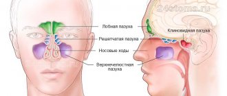
The formation of a cyst occurs due to impaired ventilation of the paranasal sinuses and the outflow of mucus, i.e. There is no physiological gas exchange and removal of mucous membrane secretions from the sinus. This leads to dysfunction of the secretory cells of the mucous membrane, which begins to grow, the gland duct is blocked and a cyst is formed in the form of a thin-walled bubble with liquid inside. The causes of the appearance of cysts are chronic inflammation of the nasal cavity, the polypous process, as well as inflammatory processes in the roots of the teeth located in close proximity to the sinuses.
Polyps of the maxillary sinus are growths of the nasal mucosa, which appear due to the presence of chronic inflammation with insufficient treatment of colds or allergic diseases, as well as in the presence of immunological changes.
Odontogenic sinusitis develops due to the penetration of infection into the maxillary sinus from an inflammatory focus in the roots of the teeth located in close proximity to the maxillary sinus. As medical practice shows, patients very often turn to an otorhinolaryngologist with symptoms of odontogenic sinusitis after dental surgery. In some patients, inflammation of the maxillary sinus was caused by an infection remaining in the tooth socket, in others, the infection penetrated into the sinus after surgical removal of the tooth, and still others complained of sinusitis due to the filling substance getting into the nasal appendages.
Symptoms of odontogenic sinusitis
Symptoms of odontogenic sinusitis, cysts and polyps
- frequent colds
- migraine-like headache
- temporary or permanent nasal congestion and difficulty breathing
- unpleasant odor from the nose and mouth
- accumulation of mucus and phlegm in the throat
- mucous, light or cloudy purulent discharge from the nose
- dysfunction of the sense of smell
Treatment
At the Federal State Budgetary Institution National Medical Research Center of Otorhinolaryngology, Federal Medical and Biological Agency of Russia, the treatment of sinusitis, cysts and polyps is carried out by specialists from the scientific and clinical department of diseases of the nose and pharynx under the guidance of a talented doctor, Ph.D. V.M. Averbukh. The Center's modern diagnostic equipment, extensive experience of doctors and the successful use of the latest developments and techniques in the field of treatment of sinusitis help not only to correctly diagnose the disease, but also to prescribe effective treatment.
The Center's specialists believe that not all types of cysts and polyps require surgical removal. As practice shows, in most cases, acute inflammation in the maxillary sinus responds well to conservative treatment - the right combination of anti-inflammatory, antibacterial and antiallergic drugs. Patients should be aware that the cyst does not resolve on its own and only in very rare cases is it evacuated from the sinus spontaneously. Only a specialist can determine the type and nature of the cyst and prescribe the correct solution to the problem. If the cyst is large and its volume puts pressure on the walls of the sinus, surgical intervention is indicated.
But before prescribing an individual course of treatment, it is necessary to conduct a thorough diagnostic examination of the patient.
Diagnostics
In the scientific and clinical department of diseases of the nose and pharynx, various modern types of diagnostics are used:
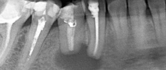
- radiography
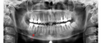
- orthopantogram (panoramic X-ray of the jaw)
— CT scan of the paranasal sinuses
— Dental CT
— Videoendoscopy of the nasal sinuses
— MRI of the paranasal sinuses
The Center’s specialists have at their disposal various techniques developed not only by the best domestic and foreign clinics, but also their own. Endoscopic surgery on the maxillary sinus, which is performed both under local anesthesia and general anesthesia, is one of the most modern techniques used in the Center. This operation requires high skill and extensive experience of the surgeon. Through the natural anastomosis of the maxillary sinus, the surgeon carefully performs endoscopic removal of the cyst. The instrument is inserted through the patient's nose without incisions or punctures. The department’s surgeons believe that this technique has no contraindications for use, does not lead to complications, and does not require a long hospital stay. However, if it was not possible to completely remove the cyst using the mentioned method, the surgeon uses a trocar to make a 4mm hole under the upper lip in the vestibule of the oral cavity.
When a foreign body or cyst is located in the bottom of the sinus, the most modern surgical technique is used - minimally invasive removal of pathological formations using infraturbinal access (through the lower nasal passage), without forming a permanent connection between the sinus and the nasal cavity and preserving the mucous membrane. The choice of the most effective surgical intervention and the method of its implementation depend on the nature, localization of the pathological process and the experience of the surgeon.
For each patient of the Center, specialists develop an individual treatment program, and the attention and caring attitude of the staff is guaranteed to everyone!